Fraud Blocker Skip to main content

Compound Eye Collection (page 7)

The compound eye, a marvel of nature's engineering, is a fascinating feature found in various insects

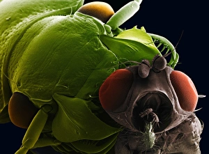
Background imageCompound Eye Collection: Bluebottle fly, SEM

Bluebottle fly, SEM
Bluebottle fly. Coloured scanning electron micrograph (SEM) of a bluebottle fly (Calliphora sp.). One of its large compound eyes (dark red) is clearly seen

Background imageCompound Eye Collection: SEM of ground beetle

SEM of ground beetle
Ground beetle. Coloured scanning electron micrograph (SEM) of a ground or carab beetle (family Carabidae) with a fly (grey) in its jaws
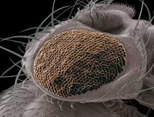
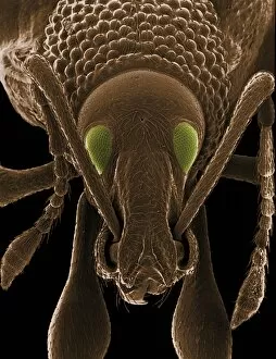
Background imageCompound Eye Collection: Coloured SEM of a weevils head

Coloured SEM of a weevils head
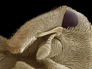
Weevil. Coloured scanning electron micrograph (SEM) of the head of a weevil. Its compound eyes (green) are seen, as well as its two elbowed (bent) antennae (centre left and right)

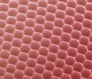
Background imageCompound Eye Collection: Compound eye of a bee, SEM

Compound eye of a bee, SEM
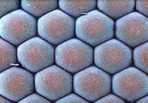
Compound eye of a bee. Coloured scanning electron micrograph (SEM) of numerous lenses making up the surface of a compound eye from a honey bee (Apis sp.)

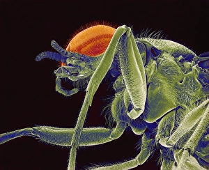
Background imageCompound Eye Collection: Fly, SEM

Fly, SEM
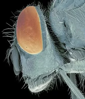
Fly. Coloured scanning electron micrograph (SEM) of the head & thorax of an unidentified fly (order Diptera). Its compound eye (yellow) and antennae (left) are seen. Magnification: x26 at 6x7cm size

Background imageCompound Eye Collection: Bluebottle fly eye, SEM

Bluebottle fly eye, SEM
Bluebottle fly (Calliphora sp.) eye, coloured scanning electron micrograph (SEM). Magnification: x4000 when printed at 10 centimetres wide

Background imageCompound Eye Collection: Hoverfly head, SEM

Hoverfly head, SEM
Hoverfly head, coloured scanning electron micrograph (SEM). Magnification: x30 when printed at 10 centimetres wide

Background imageCompound Eye Collection: Fly head, SEM

Fly head, SEM
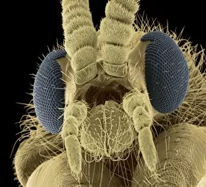
Fly head, coloured scanning electron micrograph (SEM). Close-up of the head of a fly, showing its short antennae (upper centre), which are seen between its compound eyes (blue)

Background imageCompound Eye Collection: Wasp eye, SEM

Wasp eye, SEM
Wasp eye. Coloured scanning electron micrograph (SEM) of numerous lenses, called ommatidia, that make up the compound eye of a wasp (Suborder Apocrita)

Background imageCompound Eye Collection: Male mosquito head, light micrograph

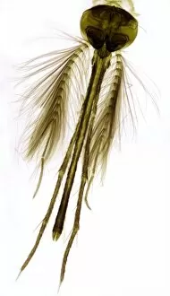
Male mosquito head, light micrograph. The head is dominated by large compound eyes. Complex sensory antennae extend from the head. A long feeding proboscis is seen between these antennae

Background imageCompound Eye Collection: Water boatman, SEM

Water boatman, SEM
Water boatman. Coloured scanning electron micrograph (SEM) showing the underside of a common water boatman (Notonecta glauca)

Background imageCompound Eye Collection: Midge head, SEM

Midge head, SEM
Midge head. Coloured scanning electron micrograph (SEM) of the head of a midge, showing its antennae (top) and one of its compound eyes (centre). Magnification: x100 when printed 10 centimetres wide

Background imageCompound Eye Collection: Clothes moth, SEM

Clothes moth, SEM
Clothes moth. Coloured scanning electron micrograph (SEM) of a common clothes moth (Tineola bisselliella) on cashmere wool fibres

Background imageCompound Eye Collection: Flour beetle, SEM

Flour beetle, SEM
Flour beetle, coloured scanning electron micrograph (SEM). Flour beetles (family Tenebrionidae) are adapted to survive in very dry environments and can withstand high amounts of radiation

Background imageCompound Eye Collection: Mosquito head, SEM

Mosquito head, SEM
Mosquito head. Coloured scanning electron micrograph (SEM) of the head of a mosquito, showing its large compound eyes. Each eye consists of many lenses (spheres) known as ommatidia

Background imageCompound Eye Collection: Bluebottle fly head, SEM

Bluebottle fly head, SEM
Bluebottle fly (Calliphora vomitoria) head, coloured scanning electron micrograph (SEM). Magnification: x35 when printed at 10 centimetres tall

Background imageCompound Eye Collection: Tropical ant head, SEM

Tropical ant head, SEM
Tropical ant head. Coloured scanning electron micrograph (SEM) of the head of a tropical ant (family Formicidae), showing one of its compound eyes (red)

Background imageCompound Eye Collection: Butterfly head, light micrograph

Butterfly head, light micrograph
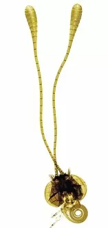
Butterfly head. Light micrograph of the head of a butterfly (bottom) with its long segmented sensory antennae (centre and top)

Background imageCompound Eye Collection: Male southern hawker dragonfly

Male southern hawker dragonfly (Aeshna cyanea) at rest. This dragonfly inhabits slow-flowing waterways, gardens and open woodland. Photographed on Studland Heath, Dorset, UK, in August

Background imageCompound Eye Collection: Compound eye, SEM

Compound eye, SEM
Compound eye. Coloured scanning electron micrograph (SEM) of an eye from a narrow-bordered bee hawk moth (Hemaris tityus), showing the numerous individual photoreceptor units (ommatidia)

Background imageCompound Eye Collection: Fruit fly eye, SEM

Fruit fly eye, SEM
Fruit fly eye. Coloured scanning electron micrograph (SEM) showing the numerous different lenses (ommatidia, spheres) that make up the compound eye of a fruit fly (Drosophila sp.)

Background imageCompound Eye Collection: Twin-lobed deerfly

Twin-lobed deerfly (Chrysops relictus). Male deerflies are nectar feeders, whereas the females bite mammals (including humans) to feed on their blood. Photographed in Italy

Background imageCompound Eye Collection: Ruddy darter dragonflies mating

Ruddy darter dragonflies mating. Close-up of a male (red) and female ruddy darter (Sympetrum sanguineum) mating on a dead seed head. Photographed in Umbria, Italy, in summer

Background imageCompound Eye Collection: Scarlet dragonfly

Scarlet dragonfly (Crocothemis erythraea) dragonfly on a plant stalk. Photographed in Italy

Background imageCompound Eye Collection: Ruddy darter dragonfly

Ruddy darter dragonfly
Ruddy darter. Close-up of a male ruddy darter (Sympetrum sanguineum) dragonfly on a plant stalk. Photographed in Umbria, Italy, in summer

Background imageCompound Eye Collection: Moth head, SEM

Moth head, SEM
Moth head, coloured scanning electron micrograph (SEM). The head is dominated by the large compound eyes (one seen, pink). Two antennae are mounted on the top of the head

Background imageCompound Eye Collection: Carpet beetle, SEM

Carpet beetle, SEM
Carpet beetle (family Dermestidae), coloured scanning electron micrograph (SEM). Two compound eyes (green) are seen, with a pair of antennae between them

Background imageCompound Eye Collection: Arthropod anatomy, artwork

Arthropod anatomy, artwork
Arthropod anatomy. Computer artwork showing the main structures and organs of a typical arthropod (legs not shown). For the labelled diagram see image: C008/8720

Background imageCompound Eye Collection: Froghopper, SEM

Froghopper, SEM
Froghopper. Coloured scanning electron micrograph (SEM) of the underside of the head of a froghopper (superfamily Cercopoidea)

Background imageCompound Eye Collection: Bedbug head, SEM

Bedbug head, SEM
Bedbug head. Coloured scanning electron micrograph (SEM) of the head of a bedbug (Cimex sp.). Antennae are seen at top, with compound eyes (purple) at the sides of its head

Background imageCompound Eye Collection: Harlequin ladybird, SEM

Harlequin ladybird, SEM
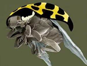
Harlequin ladybird. Coloured scanning electron micrograph (SEM) of a harlequin ladybird (Harmonia axyridis), showing its wings (grey)

Background imageCompound Eye Collection: Fungus gnat head, SEM

Fungus gnat head, SEM
Fungus gnat. Coloured scanning electron micrograph (SEM) of the head of a fungus gnat (family Sciaridae). At centre are the gnats compound eyes (brown)

Background imageCompound Eye Collection: Batocera rufomaculata beetle

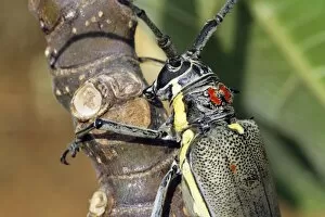
Batocera rufomaculata beetle on a plant. These beetles cause major damage to fig and mango plantations. Photographed in Israel

Background imageCompound Eye Collection: Ladybird head, SEM

Ladybird head, SEM
Ladybird head. Coloured scanning electron micrograph (SEM) of the head of a ladybird (family Coccinellidae). The compound eyes of this beetle are at left and right (blue)

Background imageCompound Eye Collection: Head of a shield bug, SEM

Head of a shield bug, SEM
Head of a shield bug (Acanthosoma sp.), coloured scanning electron micrograph (SEM). This insect is also known as a stink bug for the pungent liquid it emits if disturbed

Background imageCompound Eye Collection: Head of a crane fly, SEM

Head of a crane fly, SEM
Head of a crane fly (family Tipulidae), coloured scanning electron micrograph (SEM). The two compound eyes (purple) are either side of the head

Background imageCompound Eye Collection: Moth proboscis and eye, SEM

Moth proboscis and eye, SEM
Moth proboscis and eye, coloured scanning electron micrograph (SEM). The compound eye (orange) is at right. The spiral-shaped sucking proboscis (at centre)

Background imageCompound Eye Collection: Mutant fruit fly compound eye, SEM

Mutant fruit fly compound eye, SEM
Mutant fruit fly compound eye. Coloured scanning electron micrograph (SEM) of the abnormal compound eye of a mutant fruit fly (Drosophila melanogaster)

Background imageCompound Eye Collection: Moth eye, SEM

Moth eye, SEM
Moth eye. Coloured scanning electron micrograph (SEM) of numerous lenses making up the surface of a compound eye from a moth (order Lepidoptera). These lenses are called ommatidia

Background imageCompound Eye Collection: Weevil, SEM

Weevil, SEM
Weevil, coloured scanning electron micrograph (SEM). The weevils head is at upper left, with one of the compound eyes seen, along with both antennae

Background imageCompound Eye Collection: Weevil head, SEM

Weevil head, SEM
Weevil head, coloured scanning electron micrograph (SEM). One of the large compound eyes is at upper right, and one of the antennae is at centre

Background imageCompound Eye Collection: Polar shrimp

Polar shrimp. Close-up of the head of a polar shrimp (Lebbeus polaris), showing its compound eye (upper left) and translucent shell

Background imageCompound Eye Collection: Hoverfly head

Hoverfly head. Close-up of the head of a hoverfly (Volucella pellucens), showing its two large compound eyes and small antennae (orange) between them

Background imageCompound Eye Collection: Shrimp eyes

Shrimp eyes. Close-up of the head of a shrimp showing its large compound eyes

Background imageCompound Eye Collection: Wasp head

Wasp head. Close-up of the head of a European wasp (Vespula germanica), showing its two large compound eyes, and its antennae (black) between them

Background imageCompound Eye Collection: Head of a bee, SEM

Head of a bee, SEM
Head of a bee, coloured scanning electron micrograph (SEM). The bees compound eyes (one at upper left, red), one antenna (pointing downwards to left of eye), some of the mouthparts (below eye)


All Professionally Made to Order for Quick Shipping

The compound eye, a marvel of nature's engineering, is a fascinating feature found in various insects. Take the fruit fly for instance, as seen through the lens of an SEM Z340 / 0768. Magnified at x300 on an A4 size frame, its compound eye reveals intricate details that captivate our imagination. But it's not just the fruit fly that showcases this incredible structure. The Culex mosquito and red-barbed ant also possess compound eyes that have been meticulously examined under scanning electron micrographs (SEM). These images provide us with glimpses into their world, where vision takes on a whole new dimension. Moving on to the head of a honey bee captured by another SEM image, we witness how these tiny creatures rely heavily on their compound eyes for navigation and finding nectar-rich flowers amidst vast landscapes, and is truly remarkable how such small organisms can possess such complex visual systems. And let's not forget about other species like the hornet mimic hoverfly or even mosquitoes themselves. Their internal anatomy has been revealed through cross-sections and SEM images showcasing their feeding habits - including blood-sucking from human skin. Zooming back in to focus solely on flies, we encounter yet another stunning view of a house fly's compound eye magnified at x40 using SEM technology. This close-up view allows us to appreciate the intricate arrangement of individual lenses called ommatidia which make up this unique visual organ. With beekeeping being an essential practice worldwide, understanding the compound eye becomes crucial in managing beehives effectively. By comprehending how bees perceive their surroundings through these specialized organs, beekeepers can ensure optimal conditions for honey production and colony health. Finally returning to our initial subject - the fruit fly - we delve deeper into its microscopic world with another SEM image (Z340 / 0699).