
Cochlear Collection
"Unlocking the World of Sound: North Riding Infirmary's New Cochlear Unit" Step into a world where art meets science


All Professionally Made to Order for Quick Shipping
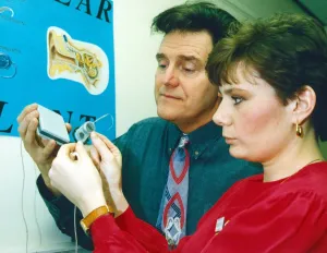
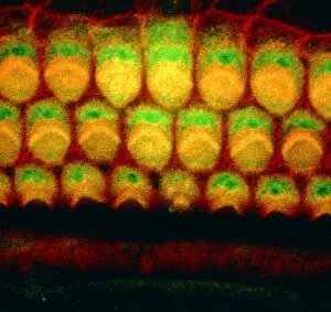
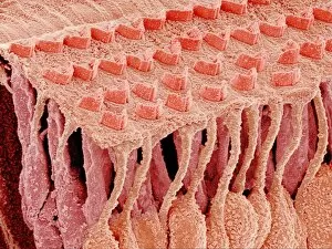
"Unlocking the World of Sound: North Riding Infirmary's New Cochlear Unit" Step into a world where art meets science, as North Riding Infirmary proudly unveils its groundbreaking unit for the hearing impaired. With a focus on female anatomy and intricate artwork, this innovative space is designed to celebrate the wonders of the cochlea. Immerse yourself in stunning visuals that depict the human ear in all its complexity. From detailed illustrations showcasing the structure of the cochlea to mesmerizing artwork depicting the hearing center, every corner tells a story of resilience and hope. Explore inner ear anatomy like never before with captivating images such as C018/6400, C018/6379, C018/6393, C018/6387, C018/6397, and C018/6402. These striking representations offer an unprecedented glimpse into our auditory system's intricacies. Marvel at microscopic wonders through scanning electron microscope (SEM) imagery capturing inner ear hair cells. Witness their delicate beauty and understand how they play a vital role in our ability to perceive sound. This new unit aims not only to provide cutting-edge medical care but also to foster understanding and appreciation for those living with hearing loss. It serves as a reminder that within each individual lies immense strength – both physically and emotionally – despite any challenges faced along their journey towards better hearing. Join us on this remarkable adventure into sound exploration as we unlock doors previously closed by deafness. Together, let us embrace diversity and empower individuals with newfound possibilities through advancements in cochlear technology.