
Cocci Collection
"Cocci: A Glimpse into the Microscopic World of Bacterial Diversity" In this captivating collection of images, we delve into the intricate world of cocci


All Professionally Made to Order for Quick Shipping
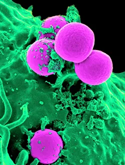
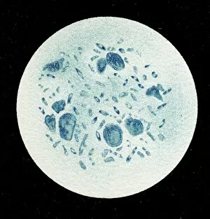
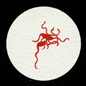
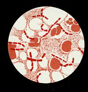
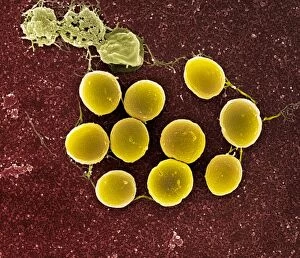
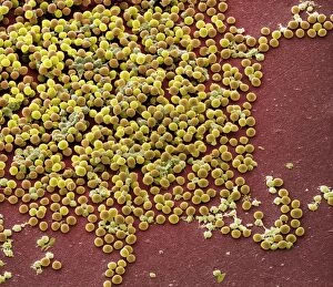
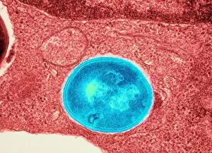
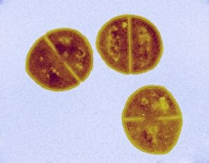
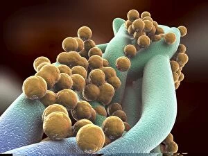
"Cocci: A Glimpse into the Microscopic World of Bacterial Diversity" In this captivating collection of images, we delve into the intricate world of cocci, a diverse group of bacteria characterized by their spherical shape. From Neutrophil engulfing MRSA to Spirillum of Lyme disease-causing bacteria, each image offers a unique glimpse into the fascinating microbial realm. The first image showcases a Neutrophil engulfing MRSA (Methicillin-resistant Staphylococcus aureus), highlighting the relentless battle between our immune system and harmful pathogens. Moving on, we encounter Cyanobacteria in all its vibrant glory through an SEM (Scanning Electron Microscope) view, reminding us of nature's incredible ability to adapt and thrive. Next up is Gonorrhoea bacteria captured using TEM (Transmission Electron Microscope), emphasizing the urgent need for effective treatments against this sexually transmitted infection. The lithographic images from 1906 take us back in time as we witness colonies of various bacterial species such as Haemophilus influenzae, Mycobacterium leprae, and Micrococcus Gonorrhoea. Continuing our journey through history, we encounter Streptococcus pneumoniae with and without bubble capsules—a visual representation that aids our understanding of how these structures contribute to pathogenicity. We then come across another colony image depicting Streptococcus Pneumoniae alone, further highlighting its significance in respiratory infections. Moving forward, we observe Spirochaetes Borrelia Recurrentis—the causative agent behind Lyme disease—within a blood sample from 1906. This lithograph serves as a reminder that even centuries ago, scientists were striving to unravel mysteries surrounding infectious diseases. Our exploration concludes with two more lithographs showcasing colonies of Vibrio cholerae—an infamous bacterium responsible for cholera outbreaks—and Clostridium tetani with spores—a bacterium that causes tetanus.