
Circulatory Collection (page 5)
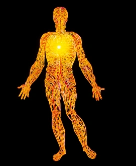
The circulatory system, also known as the cardiovascular system, is a complex network of blood vessels and organs that play a vital role in our overall health


All Professionally Made to Order for Quick Shipping
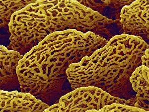
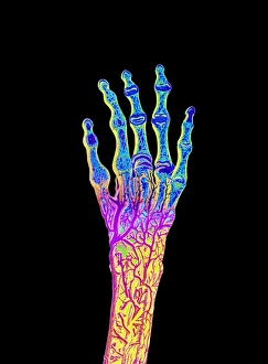
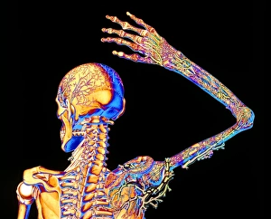
The circulatory system, also known as the cardiovascular system, is a complex network of blood vessels and organs that play a vital role in our overall health. Throughout history, artists have depicted this intricate system in their artwork, showcasing its importance and beauty. In 19th Century artwork, the neck anatomy was often portrayed with great detail. These illustrations highlighted the delicate neck vascular anatomy, emphasizing the interconnectedness of arteries and veins within this region. Such depictions not only served as artistic expressions but also provided valuable insights into medical knowledge at that time. Similarly, foot anatomy was another subject explored by illustrators during the 19th Century. Their detailed illustrations showcased various structures within the foot while highlighting its connection to circulation. This understanding became particularly relevant when studying conditions such as migraines which can be influenced by blood flow patterns. One fascinating aspect health lies in the retina's blood vessels. Scanning electron microscopy (SEM) images reveal these tiny vessels intricately woven across our retinas like delicate threads of life itself. The visualization of these blood vessels allows us to appreciate their crucial role in maintaining healthy vision. However, sometimes complications arise within our circulatory system. SEM images capturing a blood clot on plaster serve as a stark reminder of potential dangers lurking within our bodies. These powerful visuals remind us to take care and seek medical attention if necessary. The relationship between red blood cells and heart function cannot be understated either; they work hand-in-hand to ensure oxygen supply throughout our bodies efficiently. Molecular models like leptin further deepen our understanding by illustrating how hormones influence both metabolism and cardiovascular health. Platelets are another essential component involved in maintaining proper circulation; SEM images provide an up-close look at these small cell fragments responsible for clotting when injury occurs—a remarkable defense mechanism protecting us from excessive bleeding. Hand anatomy has long been studied due to its complexity and dexterity; 19th Century illustrations beautifully captured this intricacy while also highlighting the role of blood vessels within our hands.