
Ciliary Body Collection
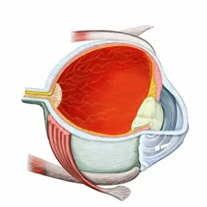
The ciliary body, located behind the iris of the eye, is a fascinating structure that plays a crucial role in our vision


All Professionally Made to Order for Quick Shipping
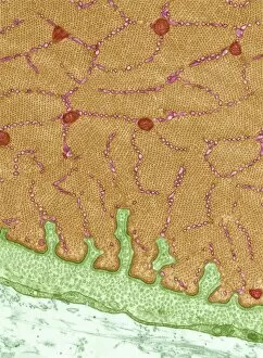
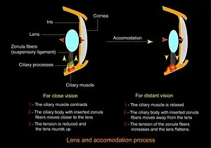
The ciliary body, located behind the iris of the eye, is a fascinating structure that plays a crucial role in our vision. When observed under scanning electron microscopy (SEM), its intricate details come to life, showcasing its complex network of tissues and cells. This eye muscle, as seen through transmission electron microscopy (TEM C014 / 1468), works tirelessly to control the shape of the lens for focusing on objects at varying distances. In another SEM image, we see the ciliary body in all its glory within the context of overall eye anatomy. This captivating snapshot from a human eye study published back in 1898 reminds us of how far our understanding has come since then. However, not all eyes are perfect. A retina affected by macular degeneration demonstrates just how delicate this vital tissue can be when it comes to clear vision. On the other hand, an image depicting advanced diabetic retinopathy serves as a stark reminder of the potential consequences if proper care is not taken. Astigmatism is yet another common condition that affects many individuals worldwide but can be corrected with corrective lenses. A conceptual image showing astigmatism and its correction provides insight into how these lenses help restore visual clarity. Returning to a conceptual cross-section view of the human eye reveals even more intricacies hidden beneath what meets our gaze every day. From cornea to optic nerve and everything in between, this image showcases various components working together seamlessly for optimal vision. Lastly, two images comparing normal and diseased retinas highlight both their similarities and differences. The former represents healthy ocular function while emphasizing just how fragile this essential part truly is; whereas the latter depicts advanced diabetic retinopathy's impact on visual health. These captivating images shed light on different aspects related to ciliary body function and overall eye anatomy – reminding us of both their beauty and vulnerability – ultimately urging us to appreciate and take care of our precious gift: sight.