
Chitin Collection
Chitin: The Intricate Beauty of Nature's Armor Step into the mesmerizing world of chitin, a remarkable substance found in various organisms


All Professionally Made to Order for Quick Shipping
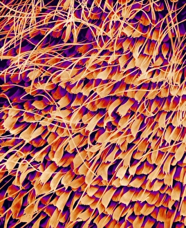
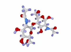
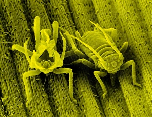
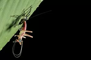
Chitin: The Intricate Beauty of Nature's Armor Step into the mesmerizing world of chitin, a remarkable substance found in various organisms. From delicate butterfly wings to the resilient exoskeletons of ticks and ants, chitin weaves its magic throughout the natural realm. Take a closer look at a butterfly wing under an electron microscope (SEM), and you'll be captivated by the intricate patterns formed by tiny overlapping scales. These scales not only provide vibrant colors but also offer protection against predators. Moving on to ticks, their exoskeletons showcase another marvel of chitin. When viewed from above, it becomes apparent how this armor shields them from harm while they latch onto their hosts for survival. Returning to butterfly wings, let us delve deeper into their enchanting beauty. Close-up images reveal the meticulous arrangement of those minuscule scales that create stunning patterns resembling works of art, and is through these structures that butterflies achieve both elegance and defense simultaneously. Butterflies are not alone in utilizing chitin's strength; ants too possess this incredible material within their anatomy. An examination of an ant anus reveals yet another example of nature's ingenuity as SEM imagery showcases the intricacies hidden beneath our naked eyes. Moths join this symphony with their own unique contribution – moth scales composed primarily of chitin. Under SEM scrutiny, these scales expose themselves as miniature wonders full of texture and detail that add grace to every fluttering motion. Venturing further into insect anatomy brings us to ant spiracles - tiny openings allowing air passage for respiration purposes. Through SEM imaging, we witness how even such minute structures rely on chitin for support and functionality. Finally, let us explore one last wonder - mosquito antennae adorned with sensory hairs made possible by none other than chitin itself. These microscopic features enable mosquitoes to detect chemical cues vital for locating potential prey or mates amidst vast landscapes.