Fraud Blocker Skip to main content

Cerebral Collection (page 3)

"Cerebral: Unlocking the Mysteries of the Mind" Delve into the intricate web of brain fibres with a DTI MRI scan C017 / 7099

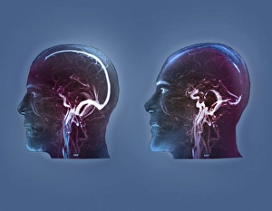
Background imageCerebral Collection: Thrombophlebitis in the brain, MRI scans

Thrombophlebitis in the brain, MRI scans
Thrombophlebitis in the brain. Coloured magnetic resonance imaging (MRI) angiograms comparing the blood vessels (white) of the brain and neck in a healthy 43-year-old (left)

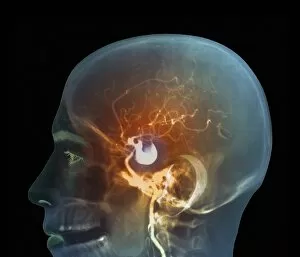
Background imageCerebral Collection: Brain aneurysm, DSA scan

Brain aneurysm, DSA scan
Brain aneurysm. Coloured digital subtraction angiogram (DSA) of the head of a 38-year-old, showing a large aneurysm (bright, centre) of the right internal carotid artery

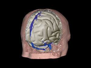
Background imageCerebral Collection: Thrombophlebitis in the brain, 3D CT scan

Thrombophlebitis in the brain, 3D CT scan
Thrombophlebitis in the brain. Coloured 3D computed tomography (CT) angiogram of the blood vessels (blue) in the brain of a 48-year-old with thrombophlebitis of the superior sagittal sinus

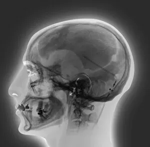
Background imageCerebral Collection: Ventricular shunt, X-ray

Ventricular shunt, X-ray
Ventricular shunt. X-ray of the head of a 52-year-old patient with hydrocephalus, showing the position of a ventricular shunt (dark lines, right) and its external valve (round, centre right)

Background imageCerebral Collection: Stroke, CT scans

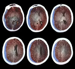
Stroke, CT scans
Stroke. Coloured computed tomography (CT) scans of sections through the brain of a patient taken 24 hours, 10 days, 25 days, and 90 days after a haemorrhagic stroke

Background imageCerebral Collection: Dementia, light micrograph

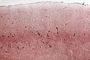
Dementia, light micrograph
Dementia. Light micrograph of a section through the brain of a patient with dementia. Dementia is mainly a disease of old age

Background imageCerebral Collection: Brain haemorrhage, CT scans C018 / 0569

Brain haemorrhage, CT scans C018 / 0569
Brain haemorrhage. Coloured computed tomography (CT) scans of the brain of a 49 year old patient with a subdural haematoma (blue)

Background imageCerebral Collection: Hydrocephalus, CT scan C018 / 0568

Hydrocephalus, CT scan C018 / 0568
Hydrocephalus. Coloured computed tomography (CT) scan of the brain of a 22 year old patient with hydrocephalus (fluid in the brain)

Background imageCerebral Collection: Thrombophlebitis, angiogram C018 / 0529

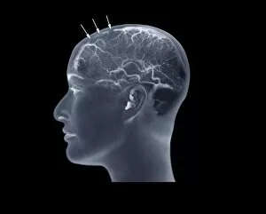
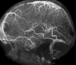
Thrombophlebitis, angiogram C018 / 0529
Thrombophlebitis. Coloured angiogram (X-ray) of the brain of a 28 year old female patient who had thrombophelibitis of the superior sagittal sinus (indicated by arrows)

Background imageCerebral Collection: Hydrocephalus, CT scan C018 / 0567

Hydrocephalus, CT scan C018 / 0567
Hydrocephalus. Coloured computed tomography (CT) scan of the brain of a 22 year old patient with hydrocephalus (fluid in the brain)

Background imageCerebral Collection: Hydrocephalus, CT scans C018 / 0566

Hydrocephalus, CT scans C018 / 0566
Hydrocephalus. Coloured computed tomography (CT) scans of the brain of a 22 year old patient with hydrocephalus (fluid in the brain)

Background imageCerebral Collection: Thrombophlebitis, angiogram C018 / 0530

Thrombophlebitis, angiogram C018 / 0530
Thrombophlebitis. Coloured angiogram (X-ray) of the brain of a 28 year old female patient who had thrombophelibitis of the superior sagittal sinus (top left)

Background imageCerebral Collection: Thrombophlebitis, angiogram C018 / 0528

Thrombophlebitis, angiogram C018 / 0528
Thrombophlebitis. Angiogram (X-ray) of the brain of a 28 year old female patient who had thrombophelibitis of the superior sagittal sinus (top left)

Background imageCerebral Collection: Corpus callosum, DTI MRI scan C017 / 7045

Corpus callosum, DTI MRI scan C017 / 7045
Corpus callosum. Coronal 3D diffusion tensor imaging (DTI) magnetic resonance imaging (MRI) scan of nerve pathways (blue) in and around the brains corpus callosum

Background imageCerebral Collection: Human brain, artwork C017 / 7399

Human brain, artwork C017 / 7399
Human brain, computer artwork

Background imageCerebral Collection: Glioblastoma brain tumour, DTI MRI scan C017 / 7052

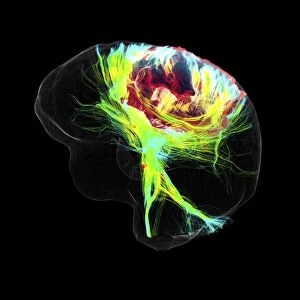
Glioblastoma brain tumour, DTI MRI scan C017 / 7052
Glioblastoma brain tumour. 3D diffusion tensor imaging (DTI) magnetic resonance imaging (MRI) scan of nerve pathways (coloured) around a glioblastoma (upper right) in the brain

Background imageCerebral Collection: Heart and brain vascular system, artwork

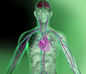
Heart and brain vascular system, artwork
Heart and brain vascular system. Artwork showing the arteries (pink) and veins (blue) of the torso, arms, neck, head and brain. The brain is at top (red) and the heart at centre

Background imageCerebral Collection: Nerve synapse, artwork C017 / 3426

Nerve synapse, artwork C017 / 3426
Nerve synapse. Computer artwork of of a junction, or synapse, between two nerve cells (neurons). As the electrical signal reaches the presynaptic end of a neuron it triggers the release of

Background imageCerebral Collection: Psychic brain, conceptual image C014 / 1480

Psychic brain, conceptual image C014 / 1480
Psychic brain, conceptual image. Artwork showing the human brain from the side, surrounded by flames, with the pineal gland shown

Background imageCerebral Collection: Psychic brain, conceptual image C014 / 1478

Psychic brain, conceptual image C014 / 1478
Psychic brain, conceptual image. Artwork of an anterior (frontal) view of the human brain. The cerebrum (the main visible area) is divided into two hemispheres

Background imageCerebral Collection: Psychic brain, conceptual image C014 / 1479

Psychic brain, conceptual image C014 / 1479
Psychic brain, conceptual image. Artwork of an anterior (frontal) view of the human brain. The cerebrum (the main visible area) is divided into two hemispheres

Background imageCerebral Collection: Brain pacemaker, artwork

Brain pacemaker, artwork
Brain pacemaker. Computer artwork showing a deep brain stimulation (DBS) electrode (right and inset) implanted in a persons brain, with a control box located on the persons chest (lower left)

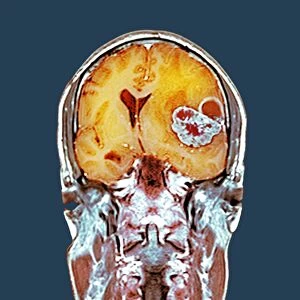
Background imageCerebral Collection: Brain cancer, MRI scan C016 / 4438

Brain cancer, MRI scan C016 / 4438
Brain cancer. Coloured coronal magnetic resonance imaging (MRI) scan through the head of a 48 year old male patient with a glioblastoma (red)

Background imageCerebral Collection: Brain cancer, MRI scan C016 / 4436

Brain cancer, MRI scan C016 / 4436
Brain cancer. Coloured coronal magnetic resonance imaging (MRI) scan through the head of a 48 year old male patient with a glioblastoma (centre right)

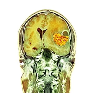
Background imageCerebral Collection: Brain cancer, MRI scan C016 / 4437

Brain cancer, MRI scan C016 / 4437
Brain cancer. Coloured coronal magnetic resonance imaging (MRI) scan through the head of a 48 year old male patient with a glioblastoma (orange)

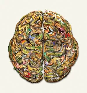
Background imageCerebral Collection: Composite brain picture

Composite brain picture
Image of a brain, composed of various flora and fauna

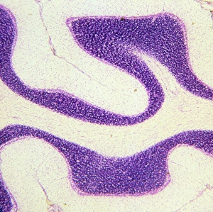
Background imageCerebral Collection: LM of section through cerebral cortex and medulla

LM of section through cerebral cortex and medulla
Cerebrum. Light micrograph of a section through the cerebrum of a human brain showing the cortex and medulla. The cerebrum is formed of two lobes (left and right)

Background imageCerebral Collection: Astronomical brain

Astronomical brain. Computer artwork of a cutaway human head revealing a brain full of stars. This could represent the use of the brain to understand the structure and origin of the universe

Background imageCerebral Collection: Brain nerve cells

Brain nerve cells
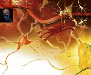
Nerve cells in the brain. Artwork showing the different types of nerve cells in the grey matter of the brain. Neurons (yellow, for example at lower right) relay nerve signals around the brain

Background imageCerebral Collection: Brain puzzle

Brain puzzle. Conceptual computer artwork of a human brain overlaid with a jigsaw puzzle pattern. This could represent research into a poorly understood brain condition such as Alzheimers disease

Background imageCerebral Collection: Human brain, occipital lobe

Human brain, occipital lobe
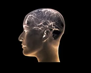
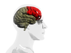
Brain, temporal lobe. Computer artwork of a human brain in profile within a glass-effect male head, showing the location of the temporal lobe (red)

Background imageCerebral Collection: Brain map

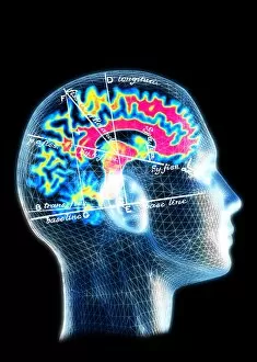
Brain map. Conceptual computer artwork of a brain with a map of anatomical features overlaid. This could represent the use of scanning techniques in medicine and experimental psychology

Background imageCerebral Collection: Space brain

Space brain

Background imageCerebral Collection: Brain

Brain. Wire-frame computer image of a side view of a healthy human brain. The front of the brain is at left. From top to centre is one of the hemispheres of the cerebrum

Background imageCerebral Collection: Brain

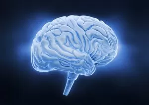
Brain. Computer artwork of a side view of a healthy human brain. At to centre is one of the hemispheres of the cerebrum, which is responsible for conscious thought, emotion and voluntary movement

Background imageCerebral Collection: Astrocyte / neuron synapse in the brain

Astrocyte / neuron synapse in the brain
Astrocyte/neuron synapse in the brain. Artwork of a nerve cell (neuron, yellow) synapse in the brain with an associated regulatory astrocyte cell (red)

Background imageCerebral Collection: Human brain, temporal lobe

Human brain, temporal lobe
Brain, brainstem. Computer artwork of a human brain in profile within a glass-effect female head, showing the location of the brainstem and central brain structures (red)

Background imageCerebral Collection: Digital brain

Digital brain. Conceptual computer artwork of a virtual human brain being examined by a magnifying glass. This could represent artificial intelligence or computer models of the brain

Background imageCerebral Collection: Virtual human

Virtual human. Conceptual computer artwork of the head of a virtual human being. This could represent artificial intelligence, or the use of computers to model the human body

Background imageCerebral Collection: Chess computer

Chess computer. Conceptual computer artwork representing a chess-playing artificial intelligence. Computers such as IBMs Deep Blue can now beat human grandmasters

Background imageCerebral Collection: Brain haemorrhage, CT scan

Brain haemorrhage, CT scan

Background imageCerebral Collection: Vision and the brain, MRI scan

Vision and the brain, MRI scan
Vision and the brain. Coloured Magnetic Resonance Imaging (MRI) scan of an axial section through the head of a 55 year old man, showing the eyes, optic nerves and brain


All Professionally Made to Order for Quick Shipping

"Cerebral: Unlocking the Mysteries of the Mind" Delve into the intricate web of brain fibres with a DTI MRI scan C017 / 7099, revealing the mesmerizing complexity of our neural pathways. Marvel at the delicate intricacy of brain blood vessels captured in a stunning 3D angiogram C007 / 1981, showcasing their vital role in nourishing our cognitive abilities. Witness synapse nerve junctions through a TEM image, where electrical signals leap across microscopic gaps to facilitate communication within our brains. Explore the challenges posed by a brain tumour as seen in an MRI scan, reminding us of both the resilience and vulnerability inherent in this remarkable organ. Discover how phantom pain after amputation is artistically depicted, shedding light on its enigmatic nature and urging empathy for those who endure it. Uncover the foundations of cognition with insights into skull anatomy and brain structure, providing essential knowledge for understanding our own consciousness. Trace back millions of years to witness primate brain evolution unfold before your eyes - a testament to nature's relentless pursuit of intelligence. Finally, observe cerebral vasculitis through an X-ray image, highlighting potential threats that can disrupt normal blood flow within this crucial system. Intriguing and awe-inspiring, "Cerebral" invites you on an extraordinary journey deep into the recesses of your mind - where wonders await at every turn.