Fraud Blocker Skip to main content

Cerebral Collection (page 2)

"Cerebral: Unlocking the Mysteries of the Mind" Delve into the intricate web of brain fibres with a DTI MRI scan C017 / 7099

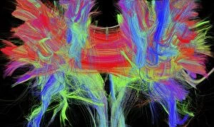
Background imageCerebral Collection: Corpus callosum, DTI MRI scan C017 / 7044

Corpus callosum, DTI MRI scan C017 / 7044
Corpus callosum. Coronal 3D diffusion tensor imaging (DTI) magnetic resonance imaging (MRI) scan of nerve pathways (coloured) in and around the brains corpus callosum

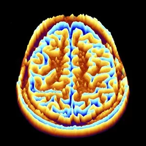
Background imageCerebral Collection: Brain scan, MRI scan, heightmap F006 / 7096

Brain scan, MRI scan, heightmap F006 / 7096
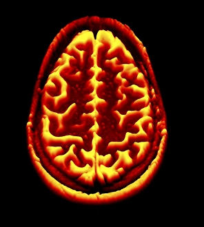
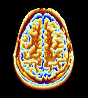
Normal brain. Coloured magnetic resonance imaging (MRI) scan of an axial section through a healthy brain, converted into a heightmap or height field

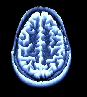
Background imageCerebral Collection: Brain scan, MRI scan, heightmap F006 / 7080

Brain scan, MRI scan, heightmap F006 / 7080
Normal brain. Coloured magnetic resonance imaging (MRI) scan of an axial section through a healthy brain, converted into a heightmap or height field
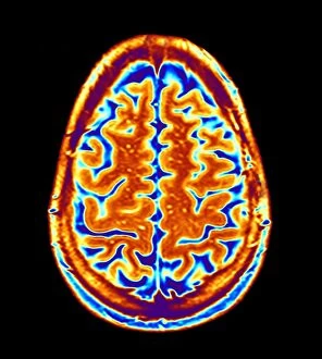
Background imageCerebral Collection: Brain scan, MRI scan F006 / 4629

Brain scan, MRI scan F006 / 4629
Normal brain. Coloured magnetic resonance imaging (MRI) scan of an axial section through a healthy brain. The front of the brain is towards top of the image

Background imageCerebral Collection: Brain scan, MRI scan F006 / 4610

Brain scan, MRI scan F006 / 4610
Normal brain. Coloured magnetic resonance imaging (MRI) scan of an axial section through a healthy brain. The front of the brain is towards top of the image

Background imageCerebral Collection: Brain scan, MRI scan F006 / 4617

Brain scan, MRI scan F006 / 4617
Normal brain. Coloured magnetic resonance imaging (MRI) scan of an axial section through a healthy brain. The front of the brain is towards top of the image

Background imageCerebral Collection: Brain scan, MRI scan F006 / 4607

Brain scan, MRI scan F006 / 4607
Normal brain. Coloured magnetic resonance imaging (MRI) scan of an axial section through a healthy brain. The front of the brain is towards top of the image

Background imageCerebral Collection: Human brain, artwork F006 / 6313

Human brain, artwork F006 / 6313
Human brain, computer artwork. The front of the brain is at bottom

Background imageCerebral Collection: Brain scan, MRI scan, heightmap F006 / 7078

Brain scan, MRI scan, heightmap F006 / 7078
Normal brain. Coloured magnetic resonance imaging (MRI) scan of an axial section through a healthy brain, converted into a heightmap or height field

Background imageCerebral Collection: Brain scan, MRI scan, heightmap F006 / 7093

Brain scan, MRI scan, heightmap F006 / 7093
Normal brain. Coloured magnetic resonance imaging (MRI) scan of an axial section through a healthy brain, converted into a heightmap or height field

Background imageCerebral Collection: Brain scan, MRI scan, heightmap F006 / 7081

Brain scan, MRI scan, heightmap F006 / 7081
Normal brain. Coloured magnetic resonance imaging (MRI) scan of an axial section through a healthy brain, converted into a heightmap or height field

Background imageCerebral Collection: Brain scan, MRI scan, heightmap F006 / 7091

Brain scan, MRI scan, heightmap F006 / 7091
Normal brain. Coloured magnetic resonance imaging (MRI) scan of an axial section through a healthy brain, converted into a heightmap or height field

Background imageCerebral Collection: Nerve cells, artwork F007 / 5514

Nerve cells, artwork F007 / 5514
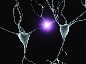
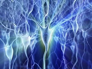
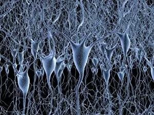
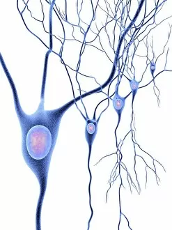
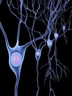
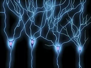
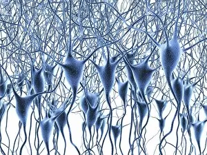
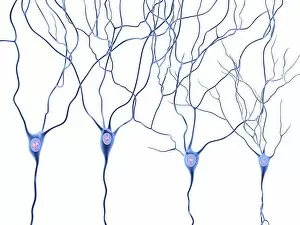
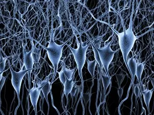
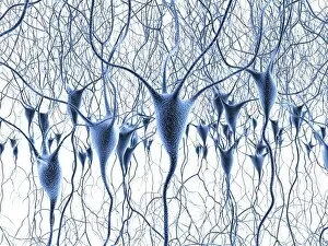
Computer artwork of nerve cells, also called neurons. Neurons are responsible for passing information around the central nervous system (CNS) and from the CNS to the rest of the body

Background imageCerebral Collection: Nerve cells, artwork F007 / 5522

Nerve cells, artwork F007 / 5522
Computer artwork of nerve cells, also called neurons. Neurons are responsible for passing information around the central nervous system (CNS) and from the CNS to the rest of the body

Background imageCerebral Collection: Nerve cells, artwork F007 / 5523

Nerve cells, artwork F007 / 5523
Computer artwork of nerve cells, also called neurons. Neurons are responsible for passing information around the central nervous system (CNS) and from the CNS to the rest of the body

Background imageCerebral Collection: Nerve cells, artwork F007 / 5526

Nerve cells, artwork F007 / 5526
Computer artwork of nerve cells, also called neurons. Neurons are responsible for passing information around the central nervous system (CNS) and from the CNS to the rest of the body

Background imageCerebral Collection: Nerve cells, artwork F007 / 5521

Nerve cells, artwork F007 / 5521
Computer artwork of nerve cells, also called neurons. Neurons are responsible for passing information around the central nervous system (CNS) and from the CNS to the rest of the body

Background imageCerebral Collection: Nerve cells, artwork F007 / 5512

Nerve cells, artwork F007 / 5512
Computer artwork of nerve cells, also called neurons. Neurons are responsible for passing information around the central nervous system (CNS) and from the CNS to the rest of the body

Background imageCerebral Collection: Nerve cells, artwork F007 / 5510

Nerve cells, artwork F007 / 5510
Computer artwork of nerve cells, also called neurons. Neurons are responsible for passing information around the central nervous system (CNS) and from the CNS to the rest of the body

Background imageCerebral Collection: Nerve cells, artwork F007 / 5530

Nerve cells, artwork F007 / 5530
Computer artwork of nerve cells, also called neurons. Neurons are responsible for passing information around the central nervous system (CNS) and from the CNS to the rest of the body

Background imageCerebral Collection: Nerve cells, artwork F007 / 5519

Nerve cells, artwork F007 / 5519
Computer artwork of firing nerve cells, also called neurons. Neurons are responsible for passing information around the central nervous system (CNS) and from the CNS to the rest of the body

Background imageCerebral Collection: Nerve cells, artwork F007 / 5518

Nerve cells, artwork F007 / 5518
Computer artwork of nerve cells, also called neurons. Neurons are responsible for passing information around the central nervous system (CNS) and from the CNS to the rest of the body

Background imageCerebral Collection: Nerve cells, artwork F007 / 5513

Nerve cells, artwork F007 / 5513
Computer artwork of nerve cells, also called neurons. Neurons are responsible for passing information around the central nervous system (CNS) and from the CNS to the rest of the body

Background imageCerebral Collection: Nerve cells, artwork F007 / 5507

Nerve cells, artwork F007 / 5507
Computer artwork of nerve cells, also called neurons. Neurons are responsible for passing information around the central nervous system (CNS) and from the CNS to the rest of the body

Background imageCerebral Collection: Nerve cells, artwork F007 / 5520

Nerve cells, artwork F007 / 5520
Computer artwork of nerve cells, also called neurons. Neurons are responsible for passing information around the central nervous system (CNS) and from the CNS to the rest of the body

Background imageCerebral Collection: Nerve cells, artwork F007 / 5525

Nerve cells, artwork F007 / 5525
Computer artwork of nerve cells, also called neurons. Neurons are responsible for passing information around the central nervous system (CNS) and from the CNS to the rest of the body

Background imageCerebral Collection: Nerve cells, artwork F007 / 5524

Nerve cells, artwork F007 / 5524
Computer artwork of nerve cells, also called neurons. Neurons are responsible for passing information around the central nervous system (CNS) and from the CNS to the rest of the body

Background imageCerebral Collection: Nerve cells, artwork F007 / 5506

Nerve cells, artwork F007 / 5506
Computer artwork of nerve cells, also called neurons. Neurons are responsible for passing information around the central nervous system (CNS) and from the CNS to the rest of the body

Background imageCerebral Collection: Nerve cells, artwork F007 / 5508

Nerve cells, artwork F007 / 5508
Computer artwork of nerve cells, also called neurons. Neurons are responsible for passing information around the central nervous system (CNS) and from the CNS to the rest of the body

Background imageCerebral Collection: Nerve cells, artwork F007 / 5515

Nerve cells, artwork F007 / 5515
Computer artwork of nerve cells, also called neurons. Neurons are responsible for passing information around the central nervous system (CNS) and from the CNS to the rest of the body

Background imageCerebral Collection: Nerve cells, artwork F007 / 5517

Nerve cells, artwork F007 / 5517
Computer artwork of nerve cells, also called neurons. Neurons are responsible for passing information around the central nervous system (CNS) and from the CNS to the rest of the body

Background imageCerebral Collection: Nerve cells, artwork F007 / 5509

Nerve cells, artwork F007 / 5509
Computer artwork of nerve cells, also called neurons. Neurons are responsible for passing information around the central nervous system (CNS) and from the CNS to the rest of the body

Background imageCerebral Collection: Nerve cells, artwork F007 / 5516

Nerve cells, artwork F007 / 5516
Computer artwork of nerve cells, also called neurons. Neurons are responsible for passing information around the central nervous system (CNS) and from the CNS to the rest of the body

Background imageCerebral Collection: Brain activity, artwork C017 / 7401

Brain activity, artwork C017 / 7401
Brain activity, computer artwork

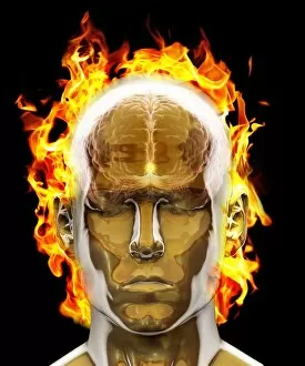
Background imageCerebral Collection: Psychic brain, conceptual image

Psychic brain, conceptual image. Artwork showing the human brain and head, surrounded in flames, with the pineal gland highlighted (bright dot in centre of forehead)

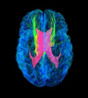
Background imageCerebral Collection: Brain fibres, DTI MRI scan C017 / 7037

Brain fibres, DTI MRI scan C017 / 7037
Brain fibres. 3D diffusion tensor imaging (DTI) magnetic resonance imaging (MRI) scan of a selection of nerve pathways (green/yellow) in the brain

Background imageCerebral Collection: Brain fibres, DTI MRI scan C017 / 7038

Brain fibres, DTI MRI scan C017 / 7038
Brain fibres. 3D diffusion tensor imaging (DTI) magnetic resonance imaging (MRI) scan of a selection of nerve pathways (green/yellow) in the brain

Background imageCerebral Collection: Brain tumour, DTI MRI scan C017 / 7057

Brain tumour, DTI MRI scan C017 / 7057
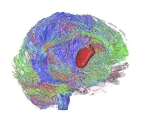
Brain tumour. 3D diffusion tensor imaging (DTI) magnetic resonance imaging (MRI) scan of nerve pathways (coloured) in a brain with a tumour (red, centre right)

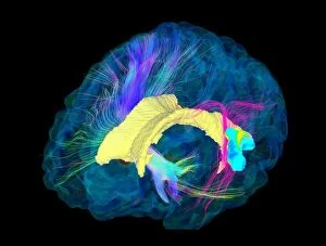
Background imageCerebral Collection: Brain fibres, DTI MRI scan C017 / 7100

Brain fibres, DTI MRI scan C017 / 7100
Brain fibres. 3D diffusion tensor imaging (DTI) magnetic resonance imaging (MRI) scan of a selection of nerve pathways (blue, pink, green) in the brain. The front of the brain is at left

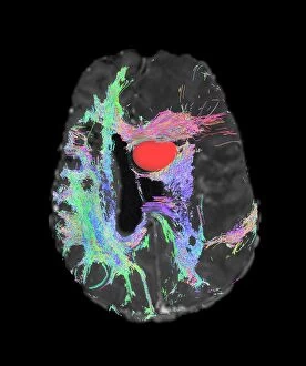
Background imageCerebral Collection: Brain tumour, DTI MRI scan C017 / 7059

Brain tumour, DTI MRI scan C017 / 7059
Brain tumour. Axial diffusion tensor imaging (DTI) magnetic resonance imaging (MRI) scan of nerve pathways (coloured) in a brain with a tumour (red, upper centre)

Background imageCerebral Collection: Brain tumour, DTI MRI scan C017 / 7058

Brain tumour, DTI MRI scan C017 / 7058
Brain tumour. 3D diffusion tensor imaging (DTI) magnetic resonance imaging (MRI) scan of nerve pathways (coloured) in a brain with a tumour (red, centre left)

Background imageCerebral Collection: Brain, neural network F007 / 0946

Brain, neural network F007 / 0946
Neural network. Computer artwork of a brain in side view, with the brains neural network represented by lines and flashes. A neural network is made up of nerve cells (neurons)

Background imageCerebral Collection: Central nervous system, artwork C017 / 7400

Central nervous system, artwork C017 / 7400
Central nervous system, computer artwork

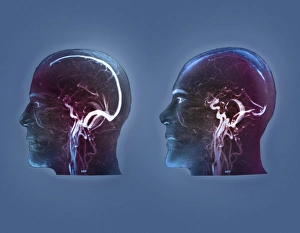
Background imageCerebral Collection: Thrombophlebitis in the brain, MRI scans

Thrombophlebitis in the brain, MRI scans
Thrombophlebitis in the brain. Coloured magnetic resonance imaging (MRI) angiograms comparing the blood vessels (white) of the brain and neck in a healthy 43-year-old (left)

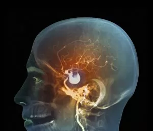
Background imageCerebral Collection: Brain aneurysm, DSA scan

Brain aneurysm, DSA scan
Brain aneurysm. Coloured digital subtraction angiogram (DSA) of the head of a 38-year-old, showing a large aneurysm (bright, centre) of the right internal carotid artery

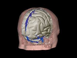
Background imageCerebral Collection: Thrombophlebitis in the brain, 3D CT scan

Thrombophlebitis in the brain, 3D CT scan
Thrombophlebitis in the brain. Coloured 3D computed tomography (CT) angiogram of the blood vessels (blue) in the brain of a 48-year-old with thrombophlebitis of the superior sagittal sinus

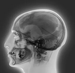
Background imageCerebral Collection: Ventricular shunt, X-ray

Ventricular shunt, X-ray
Ventricular shunt. X-ray of the head of a 52-year-old patient with hydrocephalus, showing the position of a ventricular shunt (dark lines, right) and its external valve (round, centre right)

Background imageCerebral Collection: Stroke, CT scans

Stroke, CT scans
Stroke. Coloured computed tomography (CT) scans of sections through the brain of a patient taken 24 hours, 10 days, 25 days, and 90 days after a haemorrhagic stroke


All Professionally Made to Order for Quick Shipping

"Cerebral: Unlocking the Mysteries of the Mind" Delve into the intricate web of brain fibres with a DTI MRI scan C017 / 7099, revealing the mesmerizing complexity of our neural pathways. Marvel at the delicate intricacy of brain blood vessels captured in a stunning 3D angiogram C007 / 1981, showcasing their vital role in nourishing our cognitive abilities. Witness synapse nerve junctions through a TEM image, where electrical signals leap across microscopic gaps to facilitate communication within our brains. Explore the challenges posed by a brain tumour as seen in an MRI scan, reminding us of both the resilience and vulnerability inherent in this remarkable organ. Discover how phantom pain after amputation is artistically depicted, shedding light on its enigmatic nature and urging empathy for those who endure it. Uncover the foundations of cognition with insights into skull anatomy and brain structure, providing essential knowledge for understanding our own consciousness. Trace back millions of years to witness primate brain evolution unfold before your eyes - a testament to nature's relentless pursuit of intelligence. Finally, observe cerebral vasculitis through an X-ray image, highlighting potential threats that can disrupt normal blood flow within this crucial system. Intriguing and awe-inspiring, "Cerebral" invites you on an extraordinary journey deep into the recesses of your mind - where wonders await at every turn.