Fraud Blocker Skip to main content

Cerebral Hemisphere Collection

The cerebral hemisphere, a fascinating part of the normal human brain, can be explored through various imaging techniques such as MRI and CT scans

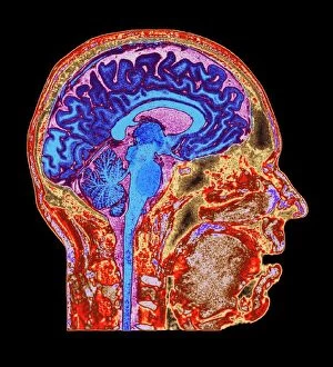
Background imageCerebral Hemisphere Collection: Normal human brain, MRI scan C016 / 8845

Normal human brain, MRI scan C016 / 8845
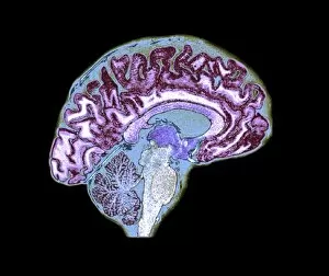
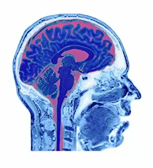
Brain. Coloured magnetic resonance imaging (MRI) scan of a sagittal section through a patients head showing a healthy human brain

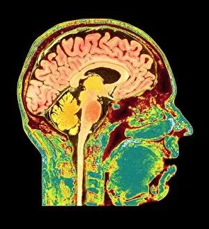
Background imageCerebral Hemisphere Collection: Normal human brain, MRI scan C016 / 8850

Normal human brain, MRI scan C016 / 8850
Brain. Coloured magnetic resonance imaging (MRI) scan of a sagittal section through a patients head showing a healthy human brain

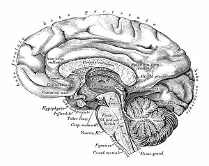
Background imageCerebral Hemisphere Collection: Human anatomy scientific illustrations: Brain side view

Human anatomy scientific illustrations: Brain side view
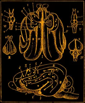
Human anatomy scientific illustrations with latin/italian labels: Brain side view

Background imageCerebral Hemisphere Collection: Stroke, MRI and 3D CT scans C016 / 6419

Stroke, MRI and 3D CT scans C016 / 6419
Stroke. Coloured composite image of a magnetic resonance imaging (MRI) scan of the brain and 2D and 3D computed tomography (CT)

Background imageCerebral Hemisphere Collection: Normal human brain, MRI scan C016 / 8843

Normal human brain, MRI scan C016 / 8843
Brain. Coloured magnetic resonance imaging (MRI) scan of a sagittal section through a healthy human brain. The highly folded area (brown) is the cerebrum, which is responsible for conscious thought

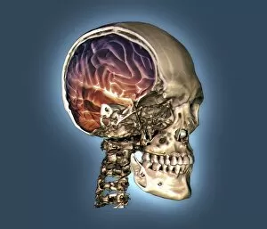
Background imageCerebral Hemisphere Collection: Normal skull and brain, 3D CT scan C016 / 6333

Normal skull and brain, 3D CT scan C016 / 6333
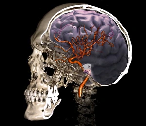
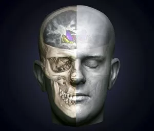
Normal skull and brain. Coloured 3D computed tomography (CT) scan of the head of a 35 year old patient. Part of the skull has been cut-away to show the brain (purple and red)

Background imageCerebral Hemisphere Collection: Normal head and neck, MRI and 3D CT scans C016 / 6337

Normal head and neck, MRI and 3D CT scans C016 / 6337
Normal head. Coloured composite image of a magnetic resonance imaging (MRI) scan of the brain and a 3D computed tomography (CT) scan of the head and neck of a 35 year old patient

Background imageCerebral Hemisphere Collection: External carotid artery, 3D CT scan C016 / 6341

External carotid artery, 3D CT scan C016 / 6341
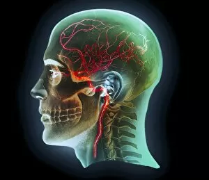
External carotid artery. Coloured 3D computed tomography (CT) angiogram (blood vessel scan) of the head showing a normal external carotid artery (red, in neck) and its branches

Background imageCerebral Hemisphere Collection: Brain anatomy

Brain anatomy
Illustration of a brain anatomy

Background imageCerebral Hemisphere Collection: Illustration of cerebral hemisphere, lower and medial surface of brain

Illustration of cerebral hemisphere, lower and medial surface of brain
Medicine: Human anatomy, cerebral hemisphere, lower and medial surface of the brain, gyri

Background imageCerebral Hemisphere Collection: Human head, MRI and 3D CT scans

Human head, MRI and 3D CT scans
Stroke. Coloured composite image of a magnetic resonance imaging (MRI) scan of the brain and 2D and 3D computed tomography (CT)

Background imageCerebral Hemisphere Collection: Human head, MRI and 3D CT scans C016 / 6407

Human head, MRI and 3D CT scans C016 / 6407
Human head. Coloured composite image of a magnetic resonance imaging (MRI) scan of the brain and a 3D computed tomography (CT) scan of the skull of a 35 year old patient

Background imageCerebral Hemisphere Collection: Human head, MRI and 3D CT scans C016 / 6417

Human head, MRI and 3D CT scans C016 / 6417
Stroke. Coloured composite image of a magnetic resonance imaging (MRI) scan of the brain and 2D and 3D computed tomography (CT)

Background imageCerebral Hemisphere Collection: Normal human brain, MRI scan C016 / 8840

Normal human brain, MRI scan C016 / 8840
Brain. Coloured magnetic resonance imaging (MRI) scan of a sagittal section through a healthy human brain. The highly folded area (yellow) is the cerebrum, which is responsible for conscious thought

Background imageCerebral Hemisphere Collection: Normal human brain, MRI scan C016 / 8848

Normal human brain, MRI scan C016 / 8848
Brain. Coloured magnetic resonance imaging (MRI) scan of a sagittal section through a patients head showing a healthy human brain

Background imageCerebral Hemisphere Collection: Human head, MRI and 3D CT scans C016 / 6397

Human head, MRI and 3D CT scans C016 / 6397
Human head. Coloured composite image of a magnetic resonance imaging (MRI) scan of the brain and a 3D computed tomography (CT) scan of the skull of a 35 year old patient

Background imageCerebral Hemisphere Collection: Normal human brain, MRI scan C016 / 8837

Normal human brain, MRI scan C016 / 8837
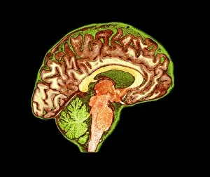
Brain. Coloured magnetic resonance imaging (MRI) scan of a sagittal section through a healthy human brain. The highly folded area (orange) is the cerebrum, which is responsible for conscious thought

Background imageCerebral Hemisphere Collection: Human head, MRI and 3D CT scans C016 / 6409

Human head, MRI and 3D CT scans C016 / 6409
Human head. Coloured composite image of a magnetic resonance imaging (MRI) scan of the brain and a 3D computed tomography (CT) scan of the skull of a 35 year old patient

Background imageCerebral Hemisphere Collection: Human head, MRI and 3D CT scans C016 / 6416

Human head, MRI and 3D CT scans C016 / 6416
Human head. Coloured composite image of a magnetic resonance imaging (MRI) scan of the brain and 2D and 3D computed tomography (CT) scans of the head and shoulders of a 38 year old patient

Background imageCerebral Hemisphere Collection: Normal human brain, MRI scan C016 / 8841

Normal human brain, MRI scan C016 / 8841
Brain. Coloured magnetic resonance imaging (MRI) scan of a sagittal section through a healthy human brain. The highly folded area (red and yellow) is the cerebrum

Background imageCerebral Hemisphere Collection: Normal human brain, MRI scan C016 / 8835

Normal human brain, MRI scan C016 / 8835
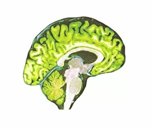
Brain. Coloured magnetic resonance imaging (MRI) scan of a sagittal section through a healthy human brain. The highly folded area across top is the cerebrum

Background imageCerebral Hemisphere Collection: Normal human brain, MRI scan C016 / 8846

Normal human brain, MRI scan C016 / 8846
Brain. Coloured magnetic resonance imaging (MRI) scan of a sagittal section through a patients head showing a healthy human brain

Background imageCerebral Hemisphere Collection: Normal human brain, MRI scan C016 / 8834

Normal human brain, MRI scan C016 / 8834
Brain. Coloured magnetic resonance imaging (MRI) scan of a sagittal section through a healthy human brain. The highly folded area (orange) is the cerebrum, which is responsible for conscious thought

Background imageCerebral Hemisphere Collection: Normal human brain, MRI scan C016 / 8849

Normal human brain, MRI scan C016 / 8849
Brain. Coloured magnetic resonance imaging (MRI) scan of a sagittal section through a patients head showing a healthy human brain

Background imageCerebral Hemisphere Collection: Normal human brain, MRI scan C016 / 8836

Normal human brain, MRI scan C016 / 8836
Brain. Coloured magnetic resonance imaging (MRI) scan of a sagittal section through a healthy human brain. The highly folded area across top is the cerebrum

Background imageCerebral Hemisphere Collection: Normal human brain, MRI scan C016 / 8844

Normal human brain, MRI scan C016 / 8844
Brain. Coloured magnetic resonance imaging (MRI) scan of a sagittal section through a patients head showing a healthy human brain

Background imageCerebral Hemisphere Collection: Internal carotid artery, 3D CT scan C016 / 6399

Internal carotid artery, 3D CT scan C016 / 6399
Internal carotid artery. Coloured 3D computed tomography (CT) angiogram (blood vessel scan) of the head showing a normal internal carotid artery (red, in neck) and its intra-cerebral branches

Background imageCerebral Hemisphere Collection: Human head, MRI and 3D CT scans C016 / 6418

Human head, MRI and 3D CT scans C016 / 6418
Human head. Coloured composite image of a magnetic resonance imaging (MRI) scan of the brain and 2D and 3D computed tomography (CT) scans of the head and shoulders of a 38 year old patient

Background imageCerebral Hemisphere Collection: Normal human brain, MRI scan C016 / 8839

Normal human brain, MRI scan C016 / 8839
Brain. Coloured magnetic resonance imaging (MRI) scan of a sagittal section through a healthy human brain. The highly folded area across top is the cerebrum

Background imageCerebral Hemisphere Collection: Internal carotid artery, 3D CT scan C016 / 6400

Internal carotid artery, 3D CT scan C016 / 6400
Internal carotid artery. Coloured 3D computed tomography (CT) angiogram (blood vessel scan) of the head showing a normal internal carotid artery (red, in neck) and its intra-cerebral branches

Background imageCerebral Hemisphere Collection: Human head, MRI and 3D CT scans C016 / 6396

Human head, MRI and 3D CT scans C016 / 6396
Human head. Coloured composite image of a magnetic resonance imaging (MRI) scan of the brain and a 3D computed tomography (CT) scan of the skull of a 35 year old patient

Background imageCerebral Hemisphere Collection: Human head, MRI and 3D CT scans C016 / 6398

Human head, MRI and 3D CT scans C016 / 6398
Human head. Coloured composite image of a magnetic resonance imaging (MRI) scan of the brain and a 3D computed tomography (CT) scan of the skull of a 35 year old patient

Background imageCerebral Hemisphere Collection: Normal human brain, MRI scan C016 / 8847

Normal human brain, MRI scan C016 / 8847
Brain. Coloured magnetic resonance imaging (MRI) scan of a sagittal section through a patients head showing a healthy human brain

Background imageCerebral Hemisphere Collection: Human head, MRI and 3D CT scans C016 / 6395

Human head, MRI and 3D CT scans C016 / 6395
Human head. Coloured composite image of a magnetic resonance imaging (MRI) scan of the brain and a 3D computed tomography (CT) scan of the skull of a 35 year old patient

Background imageCerebral Hemisphere Collection: Human head, MRI and 3D CT scans C016 / 6415

Human head, MRI and 3D CT scans C016 / 6415
Stroke. Coloured composite image of a magnetic resonance imaging (MRI) scan of the brain and 2D and 3D computed tomography (CT)

Background imageCerebral Hemisphere Collection: Normal head and neck, MRI and 3D CT scans C016 / 6338

Normal head and neck, MRI and 3D CT scans C016 / 6338
Normal head. Coloured composite image of a magnetic resonance imaging (MRI) scan of the brain and a 3D computed tomography (CT) scan of the head and neck of a 35 year old patient

Background imageCerebral Hemisphere Collection: Normal head and neck, MRI and 3D CT scans C016 / 6336

Normal head and neck, MRI and 3D CT scans C016 / 6336
Normal head. Coloured composite image of a magnetic resonance imaging (MRI) scan of the brain and a 3D computed tomography (CT) scan of the head and neck of a 35 year old patient

Background imageCerebral Hemisphere Collection: MRI scan of brain

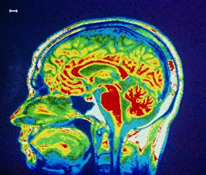
MRI scan of brain
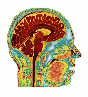
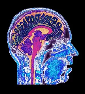
False-colour nuclear magnetic resonance (NMR) image of a mid-sagittal section through a human head, showing structures of a normal brain, airways & facial tissues

Background imageCerebral Hemisphere Collection: Thought, conceptual artwork

Thought, conceptual artwork
Thought, conceptual computer artwork

Background imageCerebral Hemisphere Collection: Human head anatomy, artwork

Human head anatomy, artwork
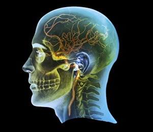
Human head anatomy. Computer artwork of the left-side of the head and neck of a human skeleton. The arteries (red lines), veins (blue lines) and nervous system (white lines) are shown


All Professionally Made to Order for Quick Shipping

The cerebral hemisphere, a fascinating part of the normal human brain, can be explored through various imaging techniques such as MRI and CT scans. In MRI scan C016 / 8845 and C016 / 8850, we get an inside look at the intricate details of this vital organ. Meanwhile, artwork depicting brain anatomy provides us with a visual representation of its complexity. Scientific illustrations showcasing the side view of the human brain (C016 / 8843) allow us to delve deeper into understanding its structure. Additionally, in the 3D CT scan C016 / 6333, we witness a comprehensive view of both the normal skull and brain. However, not all images portray healthy conditions; stroke is captured in MRI and 3D CT scans (C016 /6419), reminding us of the importance of maintaining optimal brain health. Expanding our exploration beyond just cerebral hemispheres, other scans like those for normal head and neck (C016/6337) or external carotid artery (C016/6341) provide valuable insights into related areas that contribute to overall neurological well-being. Whether it's through medical imaging or scientific illustrations, studying brain anatomy grants us invaluable knowledge about cerebral hemispheres. From their upper lateral surface to their lower and medial surfaces - every detail counts when unraveling the mysteries within our minds.