
Cell Surface Collection
The cell surface is a fascinating and complex structure that plays a crucial role in various biological processes


All Professionally Made to Order for Quick Shipping
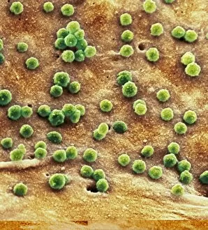
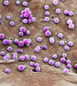
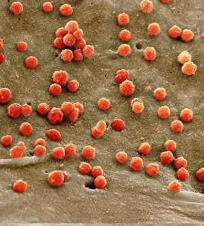
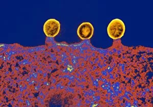
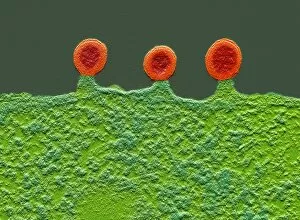
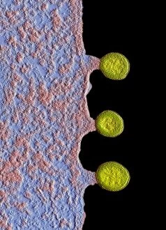
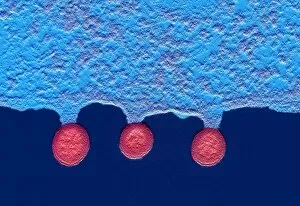
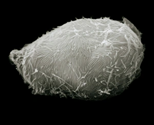
The cell surface is a fascinating and complex structure that plays a crucial role in various biological processes. One intriguing example of this is the intestinal brush border, which consists of microvilli projecting from the surface of epithelial cells lining the small intestine. These finger-like projections greatly increase the surface area available for nutrient absorption. In studying the cell surface, advanced imaging techniques such as transmission electron microscopy (TEM) have provided valuable insights. For instance, TEM images reveal the intricate details of a cell infected with HIV. SEM images labeled C014 / 0581, C014 / 0580, C014 / 0579, C017 / 8338, C017 / 8339, C017 / 8337, and C017 / 8336 showcase different views of these infected cells under high magnification. The virus hijacks host cells by binding to specific receptors on their surfaces and subsequently replicating within them. Another interesting aspect captured by TEM is the presence of budding HIV particles on mutant blood cell surfaces. These viral particles are seen emerging from infected cells as they complete their life cycle and prepare to infect new targets. Furthermore, quantum dot probes have been utilized to study the dynamics of molecules on the cell surface through artwork representations. This innovative technique allows scientists to track specific proteins or other biomolecules in real-time. Understanding how viruses interact with and manipulate cellular surfaces is essential for developing effective treatments against diseases like HIV/AIDS. By unraveling these intricate mechanisms at both structural and molecular levels using techniques like TEM and SEM, researchers continue to make significant strides towards combating viral infections and improving human health overall.