
Carotid Collection
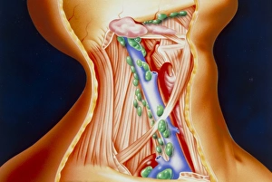
The carotid artery, a vital component of the neck vascular anatomy, holds immense significance in both historical artwork and medical illustrations


All Professionally Made to Order for Quick Shipping
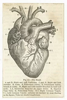
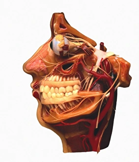
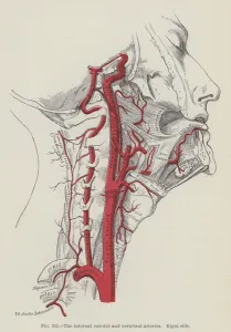
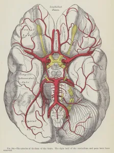
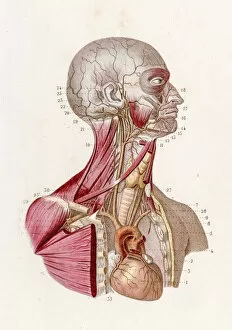
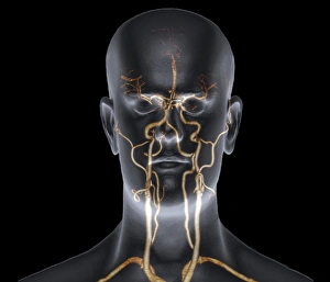
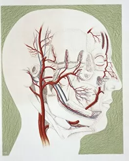
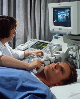
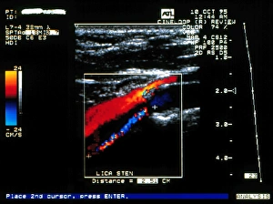
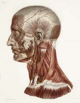
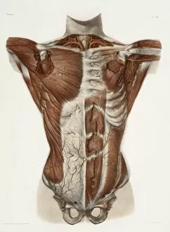
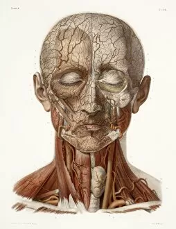
The carotid artery, a vital component of the neck vascular anatomy, holds immense significance in both historical artwork and medical illustrations. From ancient depictions to modern-day scans, the carotid artery has captivated artists and scientists alike. In Grays Anatomy, this intricate network is beautifully portrayed, showcasing its connection to the heart and its role in supplying oxygenated blood to the brain. The internal carotid and vertebral arteries are meticulously engraved on one side, revealing their importance in maintaining proper blood flow. Recherches Anatomiques Physiologiques presents a plate that highlights veins and arteries within the head. This detailed illustration offers insight into how these vessels intertwine with each other while nourishing various regions of our complex organ. An engraving depicting "The Arteries of the Base of the Brain" showcases another perspective on this crucial system. It emphasizes how these arteries form an intricate web at the foundation of our most vital organ. Vintage anatomical prints from 1912 take us back in time as they showcase not only thoracic and abdominal regions but also include details about our vascular system. These screen prints give us a glimpse into early anatomical studies that laid the groundwork for today's medical knowledge. An illustration solely dedicated to human carotid artery brings forth its prominence by highlighting its distinct features. Its path through the neck is showcased with precision, reminding us of its critical role in sustaining life-sustaining blood flow. Modern technology allows us to witness real-time images like never before; MRA scan C016/6449 reveals neck and head arteries with remarkable clarity. This non-invasive technique provides valuable insights into potential blockages or abnormalities within this essential pathway. Lastly, we see a patient undergoing doppler ultrasound—a diagnostic tool used to assess blood flow through vessels—specifically focusing on monitoring carotid artery health. This procedure enables healthcare professionals to detect any irregularities promptly while ensuring optimal blood supply to the brain.