Fraud Blocker Skip to main content

Cancerous Collection

"Cancerous: Unveiling the Battle Within" In this captivating collection of images

Background imageCancerous Collection: T lymphocytes and cancer cell, SEM C001 / 1679

T lymphocytes and cancer cell, SEM C001 / 1679
T lymphocytes and cancer cell. Coloured scanning electron micrograph (SEM) of T lymphocyte cells (green) attached to a cancer cell

Background imageCancerous Collection: Acute promyelocytic leukaemia, micrograph

Acute promyelocytic leukaemia, micrograph
Acute promyelocytic leukaemia. Light micrograph of blood cells from bone marrow in a case of acute promyelocytic leukaemia. Leukaemia is a cancer where certain blood cells form in excess

Background imageCancerous Collection: Ovarian cancer, light micrograph C015 / 7103

Ovarian cancer, light micrograph C015 / 7103
Ovary cancer. Light micrograph of a section through a Krukenberg tumour (down centre) of the ovary. This is a secondary cancer that spreads (metastasises) from the digestive system

Background imageCancerous Collection: Coloured SEM of lymphocytes attacking cancer cell

Coloured SEM of lymphocytes attacking cancer cell
Cancer cell death. Coloured Scanning Electron Micrograph (SEM) showing killer T-lymphocytes (orange) inducing a cancer cell (mauve) to undergo Programmed Cell Death (PCD)

Background imageCancerous Collection: Coloured SEM of a leukaemic white blood cell M132 / 0488

Coloured SEM of a leukaemic white blood cell M132 / 0488
Leukaemia cell. Coloured scanning electron micrograph (SEM) of a cancerous white blood cell in acute myeloid leukaemia. Leukaemia is a cancer of the blood which results in the over-production of

Background imageCancerous Collection: Polyp removal, illustration

Polyp removal, illustration. Removal of a colonic polyp with an electrical wire loop during a colonoscopy

Background imageCancerous Collection: Destruction complete d une moitie du visage par un cancer epithelial (colour litho)

Destruction complete d une moitie du visage par un cancer epithelial (colour litho)
5210424 Destruction complete d une moitie du visage par un cancer epithelial (colour litho) by French School, (19th century); Private Collection; (add.info)

Background imageCancerous Collection: The first surgical treatment of breast cancer performed under general anaesthetic by

The first surgical treatment of breast cancer performed under general anaesthetic by
CHT236348 The first surgical treatment of breast cancer performed under general anaesthetic by Seishu Hanaoka (1760-1836) in 1804 (colour litho) by Japanese School

Background imageCancerous Collection: Model of a tumour of the left ventricle, 1799 (wax)

Model of a tumour of the left ventricle, 1799 (wax)
CHT215730 Model of a tumour of the left ventricle, 1799 (wax) by French School, (18th century); Musee Dupuytren, Paris, France; (add.info)

Background imageCancerous Collection: Cancer ulcere du sein a sa derniere periode (colour litho)

Cancer ulcere du sein a sa derniere periode (colour litho)
5210386 Cancer ulcere du sein a sa derniere periode (colour litho) by French School, (19th century); Private Collection; (add.info.: Cancer ulcere du sein a sa derniere periode)

Background imageCancerous Collection: Kyste sereux cloisonne du cou, Lipome de la tete (colour litho)

Kyste sereux cloisonne du cou, Lipome de la tete (colour litho)
5210423 Kyste sereux cloisonne du cou, Lipome de la tete (colour litho) by French School, (19th century); Private Collection; (add.info.: Kyste sereux cloisonne du cou, Lipome de la tete)

Background imageCancerous Collection: Kyste areolaire de l ovaire droit a son complet developpement (colour litho)

Kyste areolaire de l ovaire droit a son complet developpement (colour litho)
5210389 Kyste areolaire de l ovaire droit a son complet developpement (colour litho) by French School, (19th century); Private Collection; (add.info)

Background imageCancerous Collection: Brain tumour, fMRI and tractography C017 / 7102

Brain tumour, fMRI and tractography C017 / 7102
Brain tumour, fMRI and tractography. Combined functional magnetic resonance imaging (fMRI, blue and green) and tractography (yellow and red) imaging of a brain with a tumour (upper left)

Background imageCancerous Collection: Secondary liver cancer, ultrasound scan C017 / 7764

Secondary liver cancer, ultrasound scan C017 / 7764
Secondary liver cancer. Ultrasound scan of a 63 year old male patients liver showing a metastatic (secondary) tumour. The cancer has metastasised (spread) from a primary cancer

Background imageCancerous Collection: Breast Cancer. Oil Painting C017 / 1273

Breast Cancer. Oil Painting C017 / 1273
Breast Cancer. Painting of a woman after breast removal surgery (mastectomy) Oil on Canvas by Nurit Shany

Background imageCancerous Collection: Brain tumour, DTI modelling C017 / 7060

Brain tumour, DTI modelling C017 / 7060
Brain tumour. Computer artwork of nerve pathways (coloured) in a brain with a tumour (white/red, upper right). The brain is seen from the front. Brain tumours can be benign or malignant (cancerous)

Background imageCancerous Collection: Brain cancer, DTI and 3D CT scans C016 / 6414

Brain cancer, DTI and 3D CT scans C016 / 6414
Brain cancer. Coloured composite image of diffusion tensor imaging (DTI) scans of the brains, and 3D computed tomography (CT) scans of the skulls, of two 35 year old patients

Background imageCancerous Collection: Cardiac lymphoma, MRI scans

Cardiac lymphoma, MRI scans
Brain tumour. Coloured magnetic resonance imaging (MRI) scans of axial sections through the chest of a 22-year-old patient, showing a tumorous lesion invading the right ventricle of the heart (large)

Background imageCancerous Collection: Throat cancer, X-rays

Throat cancer, X-rays
Throat cancer. Coloured X-rays showing the transit of a contrast medium (dense white) through the throat of a 54-year-old patient with an adenocarcinoma (cancer) of the lower oesophagus (gullet)

Background imageCancerous Collection: Chronic lymphocytic leukaemia, micrograph

Chronic lymphocytic leukaemia, micrograph
Chronic lymphocytic leukaemia. Light micrograph of blood cells sampled from a lymphatic ganglion in a case of chronic lymphocytic leukaemia, also called chronic lymphoid leukaemia (CLL)

Background imageCancerous Collection: Colon cancer, endoscopic view C016 / 8308

Colon cancer, endoscopic view C016 / 8308
Colon cancer. Endoscopic view of a malignant (cancerous) adenocarcinoma (upper right) in a patients ascending colon (large intestine)

Background imageCancerous Collection: Secondary lung cancers, X-ray

Secondary lung cancers, X-ray
Secondary lung cancers. X-ray of the chest of a 60 year old male showing malignant (cancerous) tumours (white) in the lungs

Background imageCancerous Collection: Skin cancer cell, SEM C016 / 4764

Skin cancer cell, SEM C016 / 4764
Skin cancer cell. Coloured ion-abrasion scanning electron micrograph (IA-SEM) of a melanoma cell. The large dark sphere is the cell nucleus, which contains the cells genetic information

Background imageCancerous Collection: Secondary bone cancer, X-ray C017 / 7162

Secondary bone cancer, X-ray C017 / 7162
Secondary bone cancer. X-ray of the spine of a 40 year old female with metastatic (secondary) bone cancer (bright white areas). The cancer has metastasised (spread) from a primary cancer

Background imageCancerous Collection: Cancer cell, artwork C016 / 9843

Cancer cell, artwork C016 / 9843
Cancer cell. Computer artwork of a cancerous (malignant) cell (top left) in tissue. The uneven surface and cytoplasmic projections are characteristic of cancer cells

Background imageCancerous Collection: Cancer treatment, artwork F007 / 4199

Cancer treatment, artwork F007 / 4199
Cancer treatment, conceptual image. Computer artwork representing the use of radiation in tumour treatment. Tumours are caused by the uncontrolled growth of previously normal cells

Background imageCancerous Collection: Breast cancer, artwork F006 / 7491

Breast cancer, artwork F006 / 7491
Breast cancer, computer artwork

Background imageCancerous Collection: Testicular cancer, artwork

Testicular cancer, artwork
Testicular cancer, computer artwork

Background imageCancerous Collection: Brain cancer, artwork F006 / 7482

Brain cancer, artwork F006 / 7482
Brain cancer, computer artwork

Background imageCancerous Collection: Brain cancer, artwork F006 / 7483

Brain cancer, artwork F006 / 7483
Brain cancer, computer artwork

Background imageCancerous Collection: Breast cancer, artwork F006 / 7497

Breast cancer, artwork F006 / 7497
Breast cancer, computer artwork

Background imageCancerous Collection: Intestinal cancer, artwork F007 / 6664

Intestinal cancer, artwork F007 / 6664
Intestinal cancer, computer artwork

Background imageCancerous Collection: Colon cancer, artwork F007 / 6862

Colon cancer, artwork F007 / 6862
Colon cancer, computer artwork

Background imageCancerous Collection: Colon tumor, artwork F007 / 6861

Colon tumor, artwork F007 / 6861
Colon tumor, computer artwork

Background imageCancerous Collection: Colon cancer, artwork F007 / 6876

Colon cancer, artwork F007 / 6876
Colon cancer, computer artwork

Background imageCancerous Collection: Colon cancer, artwork F008 / 0425

Colon cancer, artwork F008 / 0425
Colon cancer, computer artwork

Background imageCancerous Collection: Lung cancer, artwork F008 / 0116

Lung cancer, artwork F008 / 0116
Lung cancer, computer artwork

Background imageCancerous Collection: Colon cancer, artwork F008 / 0585

Colon cancer, artwork F008 / 0585
Colon cancer, computer artwork

Background imageCancerous Collection: Intestinal cancer, artwork F008 / 1199

Intestinal cancer, artwork F008 / 1199
Intestinal cancer, computer artwork

Background imageCancerous Collection: Intestinal cancer, artwork F008 / 1197

Intestinal cancer, artwork F008 / 1197
Intestinal cancer, computer artwork

Background imageCancerous Collection: Liver cancer, artwork F008 / 0127

Liver cancer, artwork F008 / 0127
Liver cancer, computer artwork

Background imageCancerous Collection: Intestinal cancer, artwork F008 / 1198

Intestinal cancer, artwork F008 / 1198
Intestinal cancer, computer artwork

Background imageCancerous Collection: Colon cancer, artwork F008 / 0678

Colon cancer, artwork F008 / 0678
Colon cancer, computer artwork

Background imageCancerous Collection: Colon cancer, artwork F008 / 0672

Colon cancer, artwork F008 / 0672
Colon cancer, computer artwork

Background imageCancerous Collection: Colon cancer, artwork F008 / 0306

Colon cancer, artwork F008 / 0306
Colon cancer, computer artwork

Background imageCancerous Collection: Colon cancer, artwork F008 / 0050

Colon cancer, artwork F008 / 0050
Colon cancer, computer artwork

Background imageCancerous Collection: Lung cancer, artwork F008 / 0214

Lung cancer, artwork F008 / 0214
Lung cancer, computer artwork

Background imageCancerous Collection: Colon cancer, artwork F008 / 0424

Colon cancer, artwork F008 / 0424
Colon cancer, computer artwork


All Professionally Made to Order for Quick Shipping

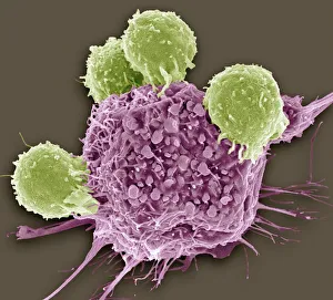
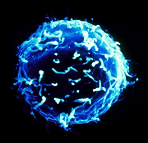
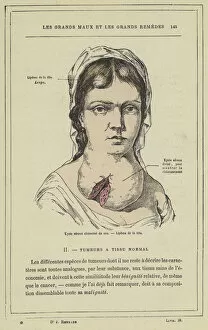
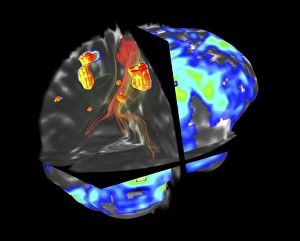
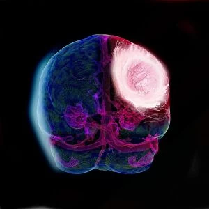
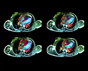
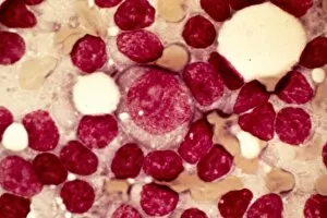
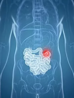
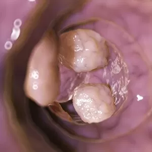
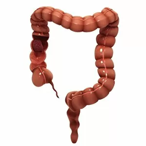
"Cancerous: Unveiling the Battle Within" In this captivating collection of images, we delve into the intricate world of cancer and witness the relentless fight between T lymphocytes and cancer cells. SEM C001/1679 reveals a mesmerizing colored SEM image showcasing lymphocytes fearlessly attacking cancer cells, symbolizing hope in the face of adversity. Acute promyelocytic leukemia takes center stage in a striking micrograph, reminding us of the urgency to combat this disease. Meanwhile, ovarian cancer unveils its hidden secrets through a light micrograph (C015/7103), urging us to unravel its mysteries for better treatment options. The battle against cancer is not limited to blood-related diseases alone; even white blood cells can succumb to its clutches. M132/0488 presents an awe-inspiring colored SEM image capturing leukaemic white blood cells under attack. Illustrating the importance of early detection and intervention, an illustration depicts polyp removal - a crucial step towards preventing further harm caused by these abnormal growths. Devastation becomes apparent as we encounter a color lithograph depicting half a face destroyed by epithelial cancer. It serves as a stark reminder that timely action is essential in combating this formidable foe. History unfolds before our eyes with an image portraying the first surgical treatment of breast cancer performed under general anesthesia. This milestone reminds us how far medical science has come in fighting against this pervasive disease. A wax model showcases a tumor within the left ventricle from 1799, highlighting both our progress and ongoing challenges in understanding and treating different types of cancers. Another poignant lithograph captures breast ulcerative cancer at its final stage – serving as motivation for continued research efforts aimed at finding effective treatments for advanced cases. Lastly, colorful lithographs depict various cysts found within different parts of our bodies – emphasizing that vigilance is key when it comes to identifying potential threats lurking beneath seemingly harmless surfaces. "Cancerous.