
Cancellous Collection
"Cancellous: Unveiling the Intricacies of Bone Tissue through SEM and MRI Scans" Exploring Osteoporotic Bone


All Professionally Made to Order for Quick Shipping
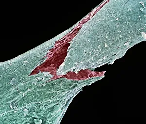
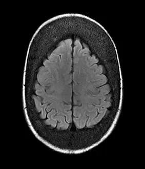
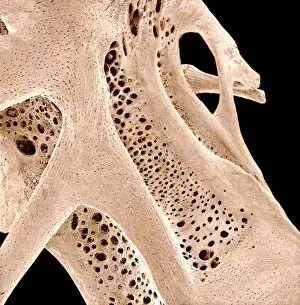
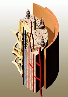
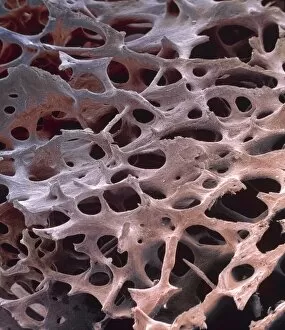
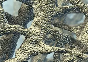
"Cancellous: Unveiling the Intricacies of Bone Tissue through SEM and MRI Scans" Exploring Osteoporotic Bone: A closer look at cancellous bone under SEM reveals its unique structure, aiding in understanding osteoporosis. Unmasking Osteoporotic Bone: SEM imaging exposes the delicate network of trabeculae within cancellous bone, offering insights into this common condition. Thickened Skull Revealed: MRI scans showcase a thickened skull, shedding light on potential underlying conditions affecting cancellous bone density. Peering Inside with MRI: Delving deeper into the mysteries bone, an MRI scan uncovers a thickened skull and prompts further investigation. Unlocking Cancellous Complexity: SEM captures intricate details bone tissue, unraveling its composition and architecture for scientific scrutiny. Diving Into Fish Bones: SEM imagery showcases fish bones' cancellous structure, providing valuable comparative data for studying human skeletal systems. The Beauty Within Fish Bones: Through stunning SEM images, we witness the mesmerizing intricacy present in fish bones' cancellous composition. Microscopic Marvels - Fish Bones Edition: Exquisite SEM visuals expose the hidden world within fish bones' cancellous structure like never before seen. Captivating Cancellations in Fish Bones: Dive into the microscopic realm as SEM highlights fascinating patterns found within fish bones' intricate cancellous makeup. Illuminating Bone Tissue Secrets: With high-resolution scanning electron microscopy (SEM), we uncover captivating details embedded within various types of bone tissues.