
Cambium Collection
"Cambium: The Lifeblood of Trees and Plants" The intricate world of cambium, the hidden hero within trees and plants


All Professionally Made to Order for Quick Shipping
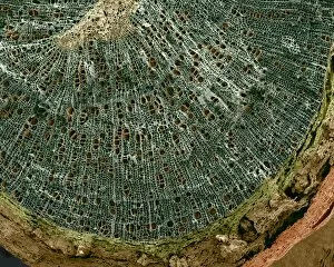
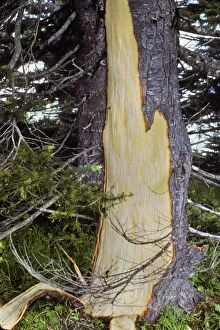
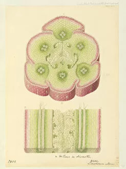
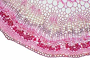
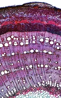
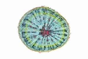
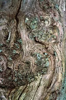
"Cambium: The Lifeblood of Trees and Plants" The intricate world of cambium, the hidden hero within trees and plants, is unveiled through fascinating glimpses captured by scientific tools. In a silver birch twig observed under a scanning electron microscope (SEM), the delicate cambium layer reveals its vital role in growth and development. Similarly, a light micrograph of a pine tree stem showcases the intricate network of cambial cells responsible for producing new wood. Nature's harmony unfolds as we delve deeper into the wonders of cambium. Bletia purpurea, an enchanting orchid species, thrives thanks to its symbiotic relationship with this remarkable tissue. Meanwhile, in Washington's Olympic National Park, evidence left behind by black bears tells an intriguing tale - these majestic creatures strip bark to feast on the thin yet nutrient-rich cambium layer. Back under the microscope lens, young pine tree stems exhibit vibrant patterns formed by actively dividing cambial cells. Lime tree stems also reveal their secrets through captivating light micrographs that showcase their own unique arrangement of this essential growth tissue. Expanding our exploration further reveals more astonishing examples: sage stems boasting intricately woven layers of cambial activity; pine stems displaying mesmerizing rings indicative of annual growth; and even pine roots demonstrating how this versatile tissue extends below ground level. In another twist showcasing nature's diversity, sunflower stems present themselves as living canvases painted with vibrant hues when examined closely under a light microscope. Each image captures the essence of life pulsating within these slender structures – all thanks to the tireless work carried out by resilient strands of cambium. Cambium truly serves as nature’s conductor orchestrating symphonies of growth and vitality throughout countless plant species worldwide. These snapshots offer us mere mortals a glimpse into its extraordinary power – reminding us that beneath every leafy canopy or towering trunk lies an intricate web where magic happens unseen but profoundly felt.