
Calculi Collection
Calculi, commonly known as urinary stones, are solid formations that can occur in various parts of the body


All Professionally Made to Order for Quick Shipping
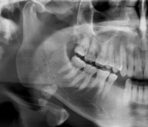
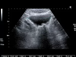
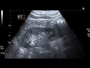
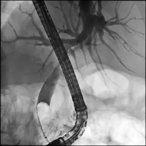
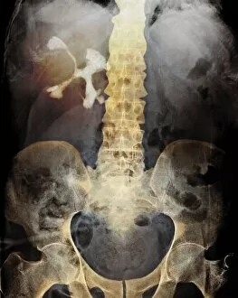
Calculi, commonly known as urinary stones, are solid formations that can occur in various parts of the body. These mineral deposits can form in different organs such as the kidneys, bladder, and even salivary glands. One type is kidney stones, which are depicted in intricate artwork. These small but painful formations develop within the kidneys and can cause severe discomfort when passing through the urinary tract. Another artwork showcases bladder stones, similar calcifications that form within the bladder itself. To diagnose these conditions accurately, medical professionals may employ imaging techniques like X-rays or ultrasound scans. In one captivating image captured by an ultrasound scan (C017 / 7262), we witness a visual representation of bladder stones inside this organ. Similarly, another scan (C017 / 7263) reveals their presence with utmost clarity. In some cases where calculi become problematic or recurrent, surgical intervention may be necessary. An illustration portrays cholecystectomy - a procedure involving the removal of the gallbladder due to gallstones formation. This conceptual image highlights how this surgery aims to alleviate pain and prevent further complications caused by these calcified structures. While calculi might seem like mere mineral formations to some observers, they hold immense significance for individuals experiencing their effects firsthand. The agony caused by kidney or bladder stones cannot be underestimated; hence understanding their nature becomes crucial for effective treatment and prevention strategies. Artworks depicting kidney stones serve as a reminder of both their beauty and potential harm they pose if left untreated (artwork C016 / 7531). By raising awareness about these conditions through visuals and medical imagery alike, we hope to encourage proactive measures towards maintaining optimal health and preventing future occurrences of calculi-related issues.