Fraud Blocker Skip to main content

Bacterial Collection (page 5)

"Bacterial: Unveiling the Microscopic World of Disease and Discovery" Step into a time capsule as we journey through history, exploring the captivating realm of bacteria

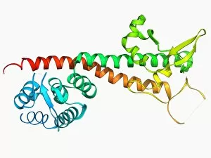
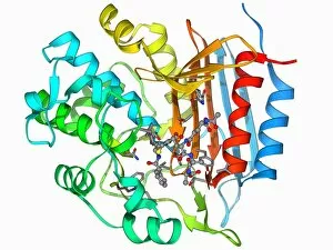
Background imageBacterial Collection: Beta-lactamase molecule F006 / 9746

Beta-lactamase molecule F006 / 9746
Beta-lactamase enzyme, molecular model. This bacterial protein provides resistance to beta-lactam antibiotics, which include penicillin

Background imageBacterial Collection: Urea channel molecule F006 / 9735

Urea channel molecule F006 / 9735
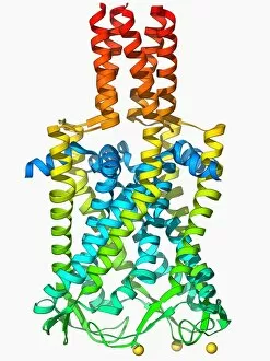
Urea channel. Molecular model of the proton-gated urea channel from the bacterium Helicobacter pylori. This channel opens in acidic environments, allowing the entry of urea into the cell

Background imageBacterial Collection: Saccharide transport protein F006 / 9722

Saccharide transport protein F006 / 9722
Saccharide transport protein. Molecular model of the phosphorylation-coupled saccharide transporter EIIC from the bacterium Bacillus cereus. EIIC is an integral membrane protein

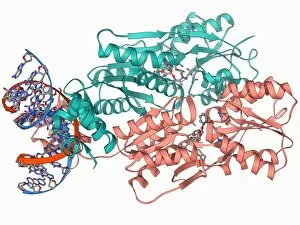
Background imageBacterial Collection: Transcription activator and DNA F006 / 9721

Transcription activator and DNA F006 / 9721
Transcription activator and DNA. Molecular model of the transcriptional activator of the multidrug efflux transporter BmrR bound to a molecule of DNA (deoxyribonucleic acid, red and blue)

Background imageBacterial Collection: Bacterial nanocompartment F006 / 9672

Bacterial nanocompartment F006 / 9672
Bacterial nanocompartment. Molecular molecule of a nanocompartment from the bacterium Thermotoga maritima. This is a shell formed from encapsulin proteins that encloses enzymes that defend the cell

Background imageBacterial Collection: Fenna-Matthews-Olson complex molecule F006 / 9673

Fenna-Matthews-Olson complex molecule F006 / 9673
Fenna-Matthews-Olson complex. Molecular model of the Fenna-Matthews-Olson complex from the green sulphur bacterium Prosthecochloris aestuarii

Background imageBacterial Collection: Bacterial biofilm enzyme F006 / 9658

Bacterial biofilm enzyme F006 / 9658
Bacterial biofilm enzyme. Molecular model of the enzyme glucansucrase from the bacterium Streptococcus mutans. This enzyme is used to form bacterial biofilms on teeth that cause dental caries

Background imageBacterial Collection: MscS ion channel protein structure F006 / 9650

MscS ion channel protein structure F006 / 9650
MscS ion channel protein structure. Molecular model of a mechanosensitive channel of small conductance (MscS) from an Escherichia coli bacterium

Background imageBacterial Collection: Streptavidin bacterial protein F006 / 9654

Streptavidin bacterial protein F006 / 9654
Streptavidin bacterial protein, molecular model. Streptavidin is a protein obtained from the bacterium Streptomyces avidinii

Background imageBacterial Collection: E coli virulence factor F006 / 9639

E coli virulence factor F006 / 9639
E. coli virulence factor. Molecular model of the beta-domain of the EspP autotransporter protein from the bacterium Escherichia coli

Background imageBacterial Collection: MscS ion channel protein structure F006 / 9626

MscS ion channel protein structure F006 / 9626
MscS ion channel protein structure. Molecular model of a mechanosensitive channel of small conductance (MscS) from an Escherichia coli bacterium

Background imageBacterial Collection: Multidrug transporter molecule F006 / 9627

Multidrug transporter molecule F006 / 9627
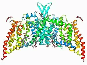
Multidrug transporter. Molecular model of the multidrug transporter Sav1866 from the bacterium Escherichia coli. This protein pumps drugs, including antibiotics, out of the bacterial cell

Background imageBacterial Collection: MscL ion channel protein structure F006 / 9624

MscL ion channel protein structure F006 / 9624
MscL ion channel protein structure. Molecular model of a mechanosensitive channel of large conductance (MscL) from a Mycobacterium tuberculosis bacterium

Background imageBacterial Collection: Rhomboid protease molecule F006 / 9621

Rhomboid protease molecule F006 / 9621
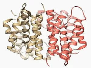
Rhomboid protease. Molecular model of the rhomboid protease enzyme GlpG from the bacterium Escherichia coli. Proteases are enzymes that break down proteins

Background imageBacterial Collection: Rhomboid protease molecule F006 / 9607

Rhomboid protease molecule F006 / 9607
Rhomboid protease. Molecular model of the rhomboid protease enzyme GlpG from the bacterium Escherichia coli. Proteases are enzymes that break down proteins

Background imageBacterial Collection: Multidrug transporter molecule F006 / 9596

Multidrug transporter molecule F006 / 9596
Multidrug transporter. Molecular model of the multidrug transporter EmrD from the bacterium Escherichia coli. This protein pumps drugs, including antibiotics, out of the bacterial cell

Background imageBacterial Collection: Respiratory complex I F006 / 9593

Respiratory complex I F006 / 9593
Respiratory complex I. Molecular model of the hydrophilic domain of respiratory complex I from the bacterium Thermus thermophilus. This enzyme is involved in energy production

Background imageBacterial Collection: RNA-induced silencing complex F006 / 9587

RNA-induced silencing complex F006 / 9587
RNA-induced silencing complex (RISC), molecular model. This complex consists of a bacterial argonaute protein (top right) bound to a small interfering RNA (siRNA) molecule (red and blue)

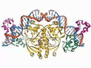
Background imageBacterial Collection: Double-stranded RNA-ribonuclease III F006 / 9585

Double-stranded RNA-ribonuclease III F006 / 9585
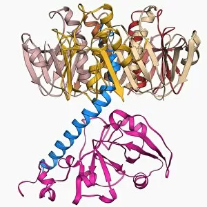
Double-stranded RNA-ribonuclease III. Molecular model of ribonuclease III (RNase III, D44N, pink and green) complexed with a double-stranded RNA (ribonucleic acid) strand (red and blue)

Background imageBacterial Collection: ATP-dependent protease molecule F006 / 9552

ATP-dependent protease molecule F006 / 9552
ATP-dependent protease. Molecular model of the bacterial enzyme HsIUV protease. Proteases are enzymes that break down proteins. HsIUV is expressed in response to cellular stress

Background imageBacterial Collection: Botulinum type B neurotoxin F006 / 9553

Botulinum type B neurotoxin F006 / 9553
Botulinum type B neurotoxin, molecular model. This powerful toxin, produced by the bacterium Clostridium botulinum, enters nerve cells and prevents release of neurotransmitters

Background imageBacterial Collection: Cholera toxin molecule F006 / 9546

Cholera toxin molecule F006 / 9546
Cholera toxin, molecular model. Cholera toxin is a protein complex secreted by the bacterium Vibrio cholerae, that causes the disease cholera

Background imageBacterial Collection: Squalene-hopene cyclase molecule F006 / 9529

Squalene-hopene cyclase molecule F006 / 9529
Squalene-hopene cyclase, molecular model. This bacterial enzyme catalyses the cyclization of squalene to hopene

Background imageBacterial Collection: LAC repressor molecule F006 / 9520

LAC repressor molecule F006 / 9520
LAC repressor. Molecular model of a LAC (lactose) repressor molecule. The LAC repressor inhibits the expression of genes that code for an enzyme which metabolizes lactose in bacteria

Background imageBacterial Collection: Staphylococcal enterotoxin C2 molecule F006 / 9506

Staphylococcal enterotoxin C2 molecule F006 / 9506
Staphylococcal enterotoxin C2. Molecular model of the C2 enterotoxin from the bacterium Staphylococcus aureus

Background imageBacterial Collection: EcoRV restriction enzyme molecule F006 / 9496

EcoRV restriction enzyme molecule F006 / 9496
EcoRV restriction enzyme. Molecular model of the type II restriction enzyme EcoRV (pink and yellow) bound to a cleaved section of DNA (deoxyribonucleic acid, red and blue)

Background imageBacterial Collection: Yersinia pestis virulence factor F006 / 9486

Yersinia pestis virulence factor F006 / 9486
Yersinia pestis virulence factor. Molecular model of the V-antigen protein, a virulence factor in Yersinia pestis, the causative agent of the plague

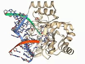
Background imageBacterial Collection: Endonuclease IV molecule F006 / 9480

Endonuclease IV molecule F006 / 9480
Endonuclease IV molecule. Molecular model of the endonuclease IV restriction enzyme EcoRV (beige) bound to a cleaved section of DNA (deoxyribonucleic acid, blue, red and green)

Background imageBacterial Collection: Outer membrane phospholipase A molecule F006 / 9469

Outer membrane phospholipase A molecule F006 / 9469
Outer membrane phospholipase A. Molecular model of the integral membrane protein, outer membrane phospholipase A from the Escherichia coli bacterium

Background imageBacterial Collection: Lactose transporter protein molecule F006 / 9466

Lactose transporter protein molecule F006 / 9466
Lactose transporter protein. Molecular model of the transmembrane transport protein lactose permease bound with a lactose homolog

Background imageBacterial Collection: Photosynthetic reaction centre F006 / 9462

Photosynthetic reaction centre F006 / 9462
Photosynthetic reaction centre. Molecular model of the photosynthetic reaction centre from the purple bacterium Rhodopseudomonas viridis

Background imageBacterial Collection: Succinate dehydrogenase enzyme F006 / 9432

Succinate dehydrogenase enzyme F006 / 9432
Succinate dehydrogenase enzyme. Molecular model of the succinate dehydrogenase (complex II) enzyme from an Escherichia coli bacterium

Background imageBacterial Collection: Diphtheria toxin, molecular model F006 / 9419

Diphtheria toxin, molecular model F006 / 9419
Diphtheria toxin, molecular model. Diphtheria is caused by the bacterium Corynebacterium diphtheriae. Symptoms include sore throat, fever and breathing difficulties

Background imageBacterial Collection: Escherichia coli heat-labile enterotoxin F006 / 9410

Escherichia coli heat-labile enterotoxin F006 / 9410
Escherichia coli heat-labile enterotoxin, molecular model. This is one of several proteins produced by pathogenic E. coli bacteria in the intestines

Background imageBacterial Collection: Gene activator protein F006 / 9406

Gene activator protein F006 / 9406
Gene activator protein. Molecular model of catabolite gene activator protein (CAP, yellow) complexed with deoxyribonucleic acid (DNA, red and blue) and RNA polymerase (green and pink)

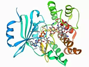
Background imageBacterial Collection: Antibiotic resistance enzyme molecule F006 / 9405

Antibiotic resistance enzyme molecule F006 / 9405
Antibiotic resistance enzyme. Molecular model of an aminoglycoside phosphotransferase enzyme. This enzyme confers resistance to aminoglycoside antibiotics

Background imageBacterial Collection: Galactoside acetyltransferase molecule F006 / 9400

Galactoside acetyltransferase molecule F006 / 9400
Galactoside acetyltransferase, molecular model. This enzyme from the bacterium Escherichia coli is involved in the lac operon

Background imageBacterial Collection: Antibiotic resistance enzyme molecule F006 / 9399

Antibiotic resistance enzyme molecule F006 / 9399
Antibiotic resistance enzyme. Molecular model of kanamycin nucleotidyltransferase (KNTase). This bacterial enzyme is responsible for resistance to a number of antibiotics

Background imageBacterial Collection: Anthrax oedema factor molecule F006 / 9393

Anthrax oedema factor molecule F006 / 9393
Anthrax oedema factor. Molecular model of oedema factor (EF) from the anthrax bacterium Bacillus antracis complexed with a calmodulin protein molecule

Background imageBacterial Collection: Anthrax lethal factor molecule F006 / 9384

Anthrax lethal factor molecule F006 / 9384
Anthrax lethal factor, molecular model. This enzyme is one of three protein components that form the anthrax toxin produced by the bacterium Bacillus anthracis

Background imageBacterial Collection: Anthrax lethal factor molecule F006 / 9379

Anthrax lethal factor molecule F006 / 9379
Anthrax lethal factor, molecular model. This enzyme is one of three protein components that form the anthrax toxin produced by the bacterium Bacillus anthracis

Background imageBacterial Collection: Multidrug efflux pump molecule F006 / 9376

Multidrug efflux pump molecule F006 / 9376
Multidrug efflux pump. Molecular model of the multidrug efflux pump AcrB from the bacterium Escherichia coli. This protein pumps drugs, including antibiotics, out of the bacterial cell

Background imageBacterial Collection: Bacterial cell wall enzyme molecule F006 / 9364

Bacterial cell wall enzyme molecule F006 / 9364
Bacterial cell wall enzyme. Molecular model of D-alanyl-D-alanine carboxypeptidase, a transpeptidase. This enzyme cross-links peptidoglycan chains in bacterial cell walls, making them rigid

Background imageBacterial Collection: Bacterial protease molecule F006 / 9340

Bacterial protease molecule F006 / 9340
Bacterial protease molecule. Computer model of a molecule of HsIUV protease complexed with its chaperone protein from a bacterium. Proteases are enzymes that break down proteins

Background imageBacterial Collection: Bacterial ribosome, molecular model F006 / 9332

Bacterial ribosome, molecular model F006 / 9332
Bacterial ribosome. Molecular model of a 30S (small) ribosomal sub-unit from the bacteria Thermus thermophilus. Ribosomes are composed of protein and RNA (ribonucleic acid)

Background imageBacterial Collection: Restriction enzyme and DNA F006 / 9315

Restriction enzyme and DNA F006 / 9315
Restriction enzyme and DNA. Molecular model showing an EcoRI endonuclease enzyme (purple and green) bound to a DNA (deoxyribonucleic acid) molecule (red and blue)

Background imageBacterial Collection: LAC repressor bound to DNA F006 / 9309

LAC repressor bound to DNA F006 / 9309
LAC repressor bound to DNA. Molecular model of a LAC (lactose) repressor molecule (pink and turquoise) interacting with bacterial DNA (deoxyribonucleic acid, red and blue)

Background imageBacterial Collection: Lumazine synthase molecule F006 / 9291

Lumazine synthase molecule F006 / 9291
Lumazine synthase molecule. Molecular model showing the structure of a lumazine synthase enzyme molecule from a Brucella abortus bacterium


All Professionally Made to Order for Quick Shipping

"Bacterial: Unveiling the Microscopic World of Disease and Discovery" Step into a time capsule as we journey through history, exploring the captivating realm of bacteria. From the haunting presence of plague doctors in 17th-century artwork to cutting-edge scientific images captured by scanning electron microscopes (SEM), prepare to be fascinated by these tiny yet formidable organisms. Witness the battle between our immune system and Methicillin-resistant Staphylococcus aureus (MRSA) as a brave neutrophil engulfs it under SEM C018/8596. Marvel at the groundbreaking Calots spinal surgery from the 19th century, which revolutionized medical practices despite lurking bacterial threats. Peering through an X-ray, tuberculosis reveals its sinister grip on human lungs while skin disorders come alive in vivid artwork, reminding us of bacteria's impact on our bodies. E. coli bacteria, magnified under SEM, showcases their intricate structures that belie their harmful potential. Travel back to Soviet Russia in 1967 with a cholera vaccination poster that symbolizes humanity's fight against bacterial epidemics. Observe salmonella bacteria under SEM; their deceptively beautiful appearance belies their ability to cause severe illness. Delve into historical diagrams depicting anthrax cultures and learn about Yersinia pestis - responsible for devastating pandemics throughout history. The tuberculosis bacterium reminds us of its long-standing presence as one of humanity's greatest adversaries. Finally, witness nature's artistic flair with spiral spore chains formed by Streptomyces bacteria – showcasing both beauty and resilience within this microscopic world. Through these glimpses into bacterial realms past and present, we gain insight into our ongoing struggle against infectious diseases. Let this exploration ignite curiosity about microbial lifeforms that shape our existence – forever reminding us how knowledge can empower us in combating these invisible foes.