Fraud Blocker Skip to main content

Bacterial Collection (page 4)

"Bacterial: Unveiling the Microscopic World of Disease and Discovery" Step into a time capsule as we journey through history, exploring the captivating realm of bacteria

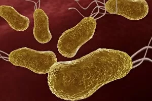
Background imageBacterial Collection: Stenotrophomonas, artwork F007 / 6768

Stenotrophomonas, artwork F007 / 6768
Stenotrophomonas bacteria, computer artwork

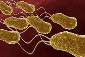
Background imageBacterial Collection: Acinetobacter, artwork F007 / 6766

Acinetobacter, artwork F007 / 6766
Acinetobacter bacteria, computer artwork

Background imageBacterial Collection: Pseudomonas, artwork F007 / 6745

Pseudomonas, artwork F007 / 6745
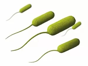
Pseudomonas bacteria, computer artwork

Background imageBacterial Collection: Klebsiella, artwork F007 / 6759

Klebsiella, artwork F007 / 6759
Klebsiella bacteria, computer artwork

Background imageBacterial Collection: MRSA bacteria, artwork F007 / 7432

MRSA bacteria, artwork F007 / 7432
MRSA (methicillin-resistant Staphylococcus aureus) bacteria, computer artwork

Background imageBacterial Collection: Enterococcus, artwork F007 / 6741

Enterococcus, artwork F007 / 6741
Enterococcus bacteria, computer artwork

Background imageBacterial Collection: Pneumocystis, artwork F007 / 6767

Pneumocystis, artwork F007 / 6767
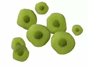
Pneumocystis bacteria, computer artwork

Background imageBacterial Collection: Morganella morganii, artwork F007 / 6765

Morganella morganii, artwork F007 / 6765
Morganella morganii bacteria, computer artwork

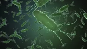
Background imageBacterial Collection: Bacteria, artwork F007 / 8494

Bacteria, artwork F007 / 8494
Bacteria, computer artwork

Background imageBacterial Collection: Glycine riboswitch molecule F007 / 9921

Glycine riboswitch molecule F007 / 9921
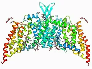
Molecular model of the bacterial glycine riboswitch. This is an RNA element that can bind the amino acid glycine. Glycine riboswitches usually consist of two metabolite-binding aptamer domains tandem

Background imageBacterial Collection: Glycine riboswitch molecule F007 / 9906

Glycine riboswitch molecule F007 / 9906
Molecular model of the bacterial glycine riboswitch. This is an RNA element that can bind the amino acid glycine. Glycine riboswitches usually consist of two metabolite-binding aptamer domains tandem

Background imageBacterial Collection: Bacteria, artwork F007 / 8491

Bacteria, artwork F007 / 8491
Bacteria, computer artwork

Background imageBacterial Collection: Bacteria, artwork F007 / 8496

Bacteria, artwork F007 / 8496
Bacteria, computer artwork

Background imageBacterial Collection: Bacteria, artwork F007 / 8495

Bacteria, artwork F007 / 8495
Bacteria, computer artwork

Background imageBacterial Collection: Bacteria shapes, artwork F007 / 9891

Bacteria shapes, artwork F007 / 9891
Computer artwork showing the three basic forms of bacteria: in the front (red) spirochetes), in the middle (green) bacilli and in the background (blue) cocci

Background imageBacterial Collection: Bacteria, artwork F007 / 8492

Bacteria, artwork F007 / 8492
Bacteria, computer artwork

Background imageBacterial Collection: Bacteria, artwork F007 / 8493

Bacteria, artwork F007 / 8493
Bacteria, computer artwork

Background imageBacterial Collection: Rod-shaped bacteria, artwork F008 / 3309

Rod-shaped bacteria, artwork F008 / 3309
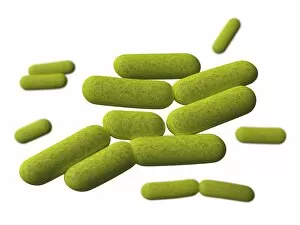
Rod-shaped bacteria, computer artwork

Background imageBacterial Collection: Rod-shaped bacteria, artwork F008 / 3310

Rod-shaped bacteria, artwork F008 / 3310
Rod-shaped bacteria, computer artwork

Background imageBacterial Collection: Bacterial intestinal infection, artwork

Bacterial intestinal infection, artwork
Bacterial intestinal infection, computer artwork

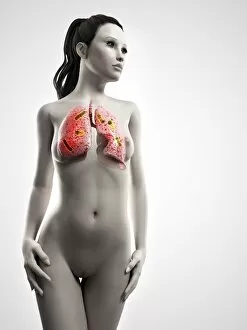
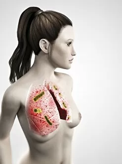
Background imageBacterial Collection: Bacterial lung infection, artwork F005 / 0557

Bacterial lung infection, artwork F005 / 0557
Bacterial lung infection, computer artwork

Background imageBacterial Collection: MRSA and dead neutrophil, SEM C018 / 8601

MRSA and dead neutrophil, SEM C018 / 8601
MRSA and dead neutrophil. Coloured scanning electron micrograph (SEM) of methicillin-resistant Staphylococcus aureus bacteria (MRSA, yellow) and a dead neutrophil white blood cell (red)

Background imageBacterial Collection: Neutrophil engulfing MRSA, SEM C018 / 8597

Neutrophil engulfing MRSA, SEM C018 / 8597
Neutrophil engulfing MRSA. Coloured scanning electron micrograph (SEM) of a neutrophil white blood cell (bottom) engulfing methicillin-resistant Staphylococcus aureus bacteria (MRSA, yellow)

Background imageBacterial Collection: E. coli Holliday junction complex C014 / 0878

E. coli Holliday junction complex C014 / 0878
E. coli Holliday junction complex. Molecular model of a RuvA protein (dark pink) in complex with a Holliday junction between homologous strands of DNA (deoxyribonucleic acid)

Background imageBacterial Collection: Staphylococcus aureus MRSA bacteria C014 / 2577

Staphylococcus aureus MRSA bacteria C014 / 2577
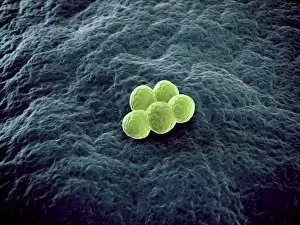
Staphylococcus aureus MRSA bacteria, computer artwork. Methicillin-resistant Staphylococcus aureus (MRSA, orange) is a Gram-positive, round (coccus) bacterium, shown here on a filamentous surface

Background imageBacterial Collection: Staphylococcus aureus MRSA bacteria C014 / 2578

Staphylococcus aureus MRSA bacteria C014 / 2578
Staphylococcus aureus MRSA bacteria, computer artwork. Methicillin-resistant Staphylococcus aureus (MRSA, orange) is a Gram-positive, round (coccus) bacterium, shown here on a filamentous surface

Background imageBacterial Collection: Iron containing protein, molecular model

Iron containing protein, molecular model
Iron containing protein. Molecular model showing the structure of a bacterial homolog of the animal iron containing protein ferritin

Background imageBacterial Collection: Endonuclease IV molecule

Endonuclease IV molecule. Molecular model of the endonuclease IV restriction enzyme EcoRV (grey) bound to a cleaved section of DNA (deoxyribonucleic acid, blue, orange and pink)

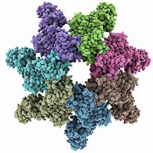
Background imageBacterial Collection: Anthrax protective antigen molecule C014 / 0886

Anthrax protective antigen molecule C014 / 0886
Anthrax protective antigen molecule. Computer model showing the structure of a molecule of protective antigen (PA) produced by anthrax (Bacillus anthracis) bacteria

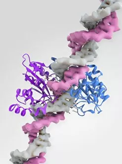
Background imageBacterial Collection: EcoRV restriction enzyme molecule C014 / 2117

EcoRV restriction enzyme molecule C014 / 2117
EcoRV restriction enzyme. Molecular model of the type II restriction enzyme EcoRV (purple and blue) bound to a DNA molecule (deoxyribonucleic acid, pink and white)

Background imageBacterial Collection: Anthrax protective antigen molecule C014 / 0865

Anthrax protective antigen molecule C014 / 0865
Anthrax protective antigen molecule. Computer model showing the structure of a molecule of protective antigen (PA) produced by anthrax (Bacillus anthracis) bacteria

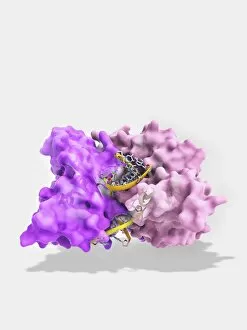
Background imageBacterial Collection: EcoRV restriction enzyme molecule C014 / 2112

EcoRV restriction enzyme molecule C014 / 2112
EcoRV restriction enzyme. Molecular model of the type II restriction enzyme EcoRV (pink) bound to a cleaved section of DNA (deoxyribonucleic acid, yellow)

Background imageBacterial Collection: Pseudomonas aeruginosa bacteria, SEM C017 / 7140

Pseudomonas aeruginosa bacteria, SEM C017 / 7140
Pseudomonas aeruginosa bacteria, coloured scanning electron micrograph (SEM). These Gram-negative rod-shaped bacteria are found in soil, water and as normal flora in the human intestine

Background imageBacterial Collection: Microbiology research F007 / 0369

Microbiology research F007 / 0369
Microbiology research

Background imageBacterial Collection: EcoRV restriction enzyme molecule C014 / 2114

EcoRV restriction enzyme molecule C014 / 2114
EcoRV restriction enzyme. Molecular model of the type II restriction enzyme EcoRV (white and gold) bound to a cleaved section of DNA (deoxyribonucleic acid, orange and yellow)

Background imageBacterial Collection: Staphylococcus aureus bacteria, SEM C017 / 7138

Staphylococcus aureus bacteria, SEM C017 / 7138
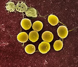
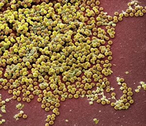
Staphylococcus aureus bacteria, coloured scanning electron micrograph (SEM). These Gram-positive coccus (round) bacteria are found on the skin and mucous membranes of humans and many animals

Background imageBacterial Collection: EcoRV restriction enzyme molecule C014 / 2116

EcoRV restriction enzyme molecule C014 / 2116
EcoRV restriction enzyme. Molecular model of the type II restriction enzyme EcoRV (purple and blue) bound to a DNA molecule (deoxyribonucleic acid, pink and white)

Background imageBacterial Collection: EcoRV restriction enzyme molecule C014 / 2115

EcoRV restriction enzyme molecule C014 / 2115
EcoRV restriction enzyme. Molecular model of the type II restriction enzyme EcoRV (purple and blue) bound to a DNA molecule (deoxyribonucleic acid, pink and white)

Background imageBacterial Collection: Staphylococcus aureus bacteria, SEM C017 / 7136

Staphylococcus aureus bacteria, SEM C017 / 7136
Staphylococcus aureus bacteria, coloured scanning electron micrograph (SEM). These Gram-positive coccus (round) bacteria are found on the skin and mucous membranes of humans and many animals

Background imageBacterial Collection: Palmitoyloleoyl PE lipid bilayer F006 / 9780

Palmitoyloleoyl PE lipid bilayer F006 / 9780
Palmitoyloleoyl PE (POPE) lipid bilayer, computer simulation. This is a model of the phospholipid bilayer of the inner bacterial membrane

Background imageBacterial Collection: Microbiology research F006 / 8583

Microbiology research F006 / 8583
Microbiology research

Background imageBacterial Collection: Bacterial alpha-hemolysin toxin F006 / 9771

Bacterial alpha-hemolysin toxin F006 / 9771
Bacterial alpha-hemolysin toxin, molecular model. This toxin is secreted by the bacterium Staphylococcus aureus. It is an example of a pore-forming toxin

Background imageBacterial Collection: Phosphofructokinase bacterial enzyme F006 / 9762

Phosphofructokinase bacterial enzyme F006 / 9762
Phosphofructokinase enzyme, molecular model. This enzyme, from the bacterium Bacillus stearothermophilus, is involved in regulating the process of releasing energy from glucose

Background imageBacterial Collection: Multidrug efflux pump molecule F006 / 9748

Multidrug efflux pump molecule F006 / 9748
Multidrug efflux pump. Molecular model of the multidrug efflux pump AcrB from the bacterium Escherichia coli transporting two doxorubicin molecules

Background imageBacterial Collection: Beta-lactamase molecule F006 / 9746

Beta-lactamase molecule F006 / 9746
Beta-lactamase enzyme, molecular model. This bacterial protein provides resistance to beta-lactam antibiotics, which include penicillin

Background imageBacterial Collection: Urea channel molecule F006 / 9735

Urea channel molecule F006 / 9735
Urea channel. Molecular model of the proton-gated urea channel from the bacterium Helicobacter pylori. This channel opens in acidic environments, allowing the entry of urea into the cell

Background imageBacterial Collection: Saccharide transport protein F006 / 9722

Saccharide transport protein F006 / 9722
Saccharide transport protein. Molecular model of the phosphorylation-coupled saccharide transporter EIIC from the bacterium Bacillus cereus. EIIC is an integral membrane protein

Background imageBacterial Collection: Transcription activator and DNA F006 / 9721

Transcription activator and DNA F006 / 9721
Transcription activator and DNA. Molecular model of the transcriptional activator of the multidrug efflux transporter BmrR bound to a molecule of DNA (deoxyribonucleic acid, red and blue)


All Professionally Made to Order for Quick Shipping

"Bacterial: Unveiling the Microscopic World of Disease and Discovery" Step into a time capsule as we journey through history, exploring the captivating realm of bacteria. From the haunting presence of plague doctors in 17th-century artwork to cutting-edge scientific images captured by scanning electron microscopes (SEM), prepare to be fascinated by these tiny yet formidable organisms. Witness the battle between our immune system and Methicillin-resistant Staphylococcus aureus (MRSA) as a brave neutrophil engulfs it under SEM C018/8596. Marvel at the groundbreaking Calots spinal surgery from the 19th century, which revolutionized medical practices despite lurking bacterial threats. Peering through an X-ray, tuberculosis reveals its sinister grip on human lungs while skin disorders come alive in vivid artwork, reminding us of bacteria's impact on our bodies. E. coli bacteria, magnified under SEM, showcases their intricate structures that belie their harmful potential. Travel back to Soviet Russia in 1967 with a cholera vaccination poster that symbolizes humanity's fight against bacterial epidemics. Observe salmonella bacteria under SEM; their deceptively beautiful appearance belies their ability to cause severe illness. Delve into historical diagrams depicting anthrax cultures and learn about Yersinia pestis - responsible for devastating pandemics throughout history. The tuberculosis bacterium reminds us of its long-standing presence as one of humanity's greatest adversaries. Finally, witness nature's artistic flair with spiral spore chains formed by Streptomyces bacteria – showcasing both beauty and resilience within this microscopic world. Through these glimpses into bacterial realms past and present, we gain insight into our ongoing struggle against infectious diseases. Let this exploration ignite curiosity about microbial lifeforms that shape our existence – forever reminding us how knowledge can empower us in combating these invisible foes.