Fraud Blocker Skip to main content

Back Bone Collection (page 3)

The backbone, also known as the spine, is a remarkable structure that serves as the foundation of our body's support system

Background imageBack Bone Collection: Skull

Skull. Computer artwork of the bones and musculature of a human head. The skull consists of the cranium, which encloses the brain, and the jaw. Embedded in the jaw are the teeth

Background imageBack Bone Collection: Spine

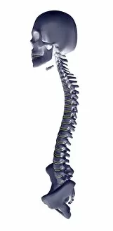
Spine. Computer artwork of a healthy spine. At top is the skull and at bottom are the bones of the pelvis. The spine is a column of 33 cylindrical bones called vertebrae

Background imageBack Bone Collection: Spine

Spine. Computer artwork of a healthy spine seen from the side. At top is the skull and at bottom are the bones of the pelvis

Background imageBack Bone Collection: Illustration of the human spine

Illustration of the human spine
Human spine. Illustration of the human spine (also called the backbone or vertebral column) seen from the side, with the rear part of the bones at left

Background imageBack Bone Collection: Spinal column

Spinal column. Historical artwork of the bones of the human spine and pelvis, seen from the front. The backbone (vertical)

Background imageBack Bone Collection: Spinal vertebra

Spinal vertebra. Historical artwork of a vertebra (spinal bone) seen from the side. The rear of the spine is at right. This is a lumbar vertebra, one from the lower back

Background imageBack Bone Collection: Spinal vertebrae

Spinal vertebrae. Historical artwork of vertebrae (spinal bones) seen from the side. The rear of the spine is at right. Four vertebrae are seen here

Background imageBack Bone Collection: Spinal bones and ligaments

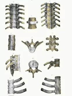
Spinal bones and ligaments. Historical anatomical artwork of thoracic (upper) spinal bones (yellow, vertebrae) and ligaments (white)

Background imageBack Bone Collection: Human body postures, historical artwork

Human body postures, historical artwork
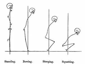
Human postures, historical artwork. The images show an outline of the shape of the body in four postures; standing, bowing, stooping and squatting

Background imageBack Bone Collection: Arthritis of the spine, X-ray

Arthritis of the spine, X-ray

Background imageBack Bone Collection: Arthritis of the back, X-ray

Arthritis of the back, X-ray
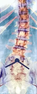
Arthritis of the back, coloured X-ray. Lumbar arthritis caused by chondrocalcinosis, a condition where crystals of calcium pyrophosphate are deposited in soft tissues

Background imageBack Bone Collection: Osteoporosis of the back, X-ray

Osteoporosis of the back, X-ray
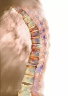
Osteoporosis of the back, coloured X-ray. Osteoporosis is a condition in which bone density is reduced, weakening the bones and making them susceptible to fractures

Background imageBack Bone Collection: Pinned curved spine, X-ray

Pinned curved spine, X-ray
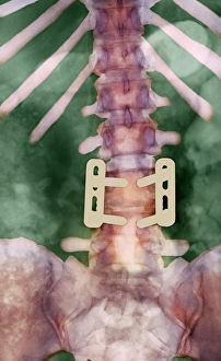
Pinned curved spine. Coloured X-ray of bones of the spine and pelvis of a 49 year old woman showing two screws (blue) pinning the lower lumbar vertebra (L5) to the sacrum of the pelvis

Background imageBack Bone Collection: Scoliosis spine deformity, X-ray

Scoliosis spine deformity, X-ray
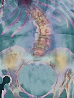
Scoliosis sideways deformity of the spine of a woman, coloured X-ray. The spine (upper centre) is severely bent. The deformity affected the patients posture

Background imageBack Bone Collection: Osteoporosis of the spine, CT scan

Osteoporosis of the spine, CT scan
Osteoporosis of the spine. Coloured 3D computed tomography (CT) scan of the twisted lumbar vertebrae of a woman with osteoporosis

Background imageBack Bone Collection: Spine degeneration, MRI scan

Spine degeneration, MRI scan
Spine degeneration. Coloured magnetic resonance imaging (MRI) scan of the lumbar (lower) region of a patients back showing spinal degeneration. The base of the spine is at lower right

Background imageBack Bone Collection: Pinned vertebrae, X-ray

Pinned vertebrae, X-ray
Pinned vertebrae. Coloured X-ray of pins (beige) joining two of the vertebrae (pink) of a 29 year old woman together to immobilise them. Vertebrae are the box-like bones that make up the spine

Background imageBack Bone Collection: Slipped disc, CT scan

Slipped disc, CT scan
Slipped disc. Coloured computed tomography (CT) scan of an axial section through an inter- vertebral disc (orange) showing herniation

Background imageBack Bone Collection: Osteoporosis of spine, X-ray

Osteoporosis of spine, X-ray
Osteoporosis. Coloured X-ray of a rear view of the lumbar (lower) spine of an elderly woman showing scoliosis (sideways curvature) of the spine caused by osteoporosis

Background imageBack Bone Collection: Osteoarthritis of neck, X-ray

Osteoarthritis of neck, X-ray
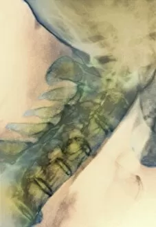
Osteoarthritis of the neck. Coloured X-ray of a side view of the neck of an elderly man, showing degenerative osteoarthritis of the cervical bones of the neck. Also known as cervical spondylosis

Background imageBack Bone Collection: Bony spurs in neck, X-ray

Bony spurs in neck, X-ray
Bony spurs in neck, coloured X-ray. Bony spurs (osteophytes, left) grow from the vertebrae as a normal part of the aging process

Background imageBack Bone Collection: Degenerative disc disease, 3D CT scan

Degenerative disc disease, 3D CT scan
Degenerative disc disease. Coloured 3D computed tomography (CT) scan of the lumbosacral spine of a 74 year old patient with degenerative disc disease


All Professionally Made to Order for Quick Shipping

The backbone, also known as the spine, is a remarkable structure that serves as the foundation of our body's support system. Just like a diagram of the human brain and spinal column reveals the intricate connection between these vital components, our backbone plays a crucial role in maintaining our overall health. Similar to the DNA molecule that carries genetic information within every cell, our spine holds essential information about our physical well-being. It acts as a central hub for communication between different parts of the body through its network of nerves. Centuries ago, medical pioneers like Calots introduced groundbreaking techniques such as spinal surgery to address issues related to this complex structure. Their efforts paved the way for advancements in understanding and treating conditions affecting not only bones but also other aspects connected to it. Artwork depicting DNA molecules reminds us that even at a microscopic level, there is an inherent link between genetics and spine health. The neck and shoulder arteries visible on X-rays demonstrate how blood flow nourishes this critical area, ensuring optimal functioning. X-ray images showcasing normal spines give us insight into bone structure from a back view perspective. These visuals allow us to appreciate how each vertebra contributes to forming this sturdy yet flexible framework supporting our entire body. However, sometimes challenges arise such as slipped discs which can cause discomfort or pain. Artistic representations help illustrate these conditions visually while MRI scans provide detailed insights into their impact on normal torso structures. Ultimately, it is important not only to understand anatomy but also take care of our backbones by adopting healthy habits like regular exercise and proper posture. By appreciating both its complexity and resilience, we can ensure that this integral part of ourselves remains strong throughout life's journey.