
Back Bone Collection
The backbone, also known as the spine, is a remarkable structure that serves as the foundation of our body's support system


All Professionally Made to Order for Quick Shipping
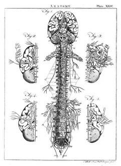
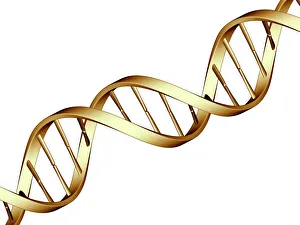
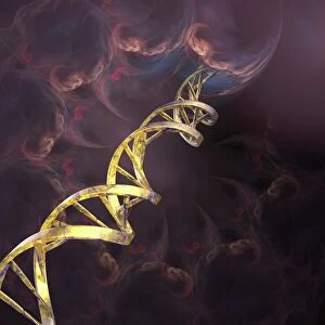
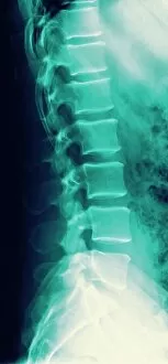
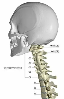
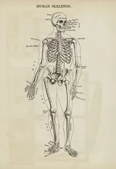
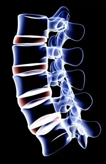
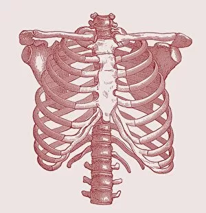
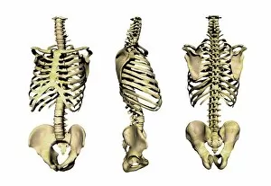
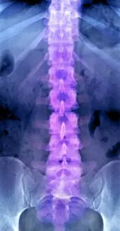
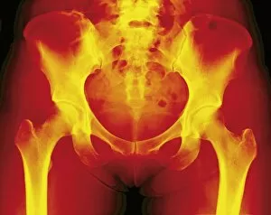
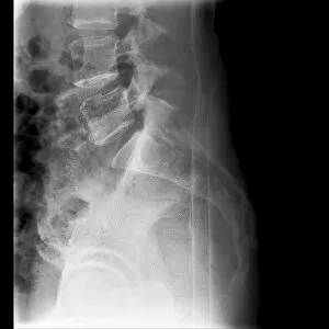
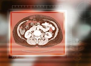
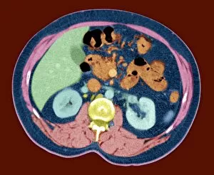
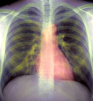
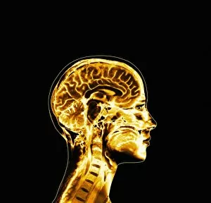
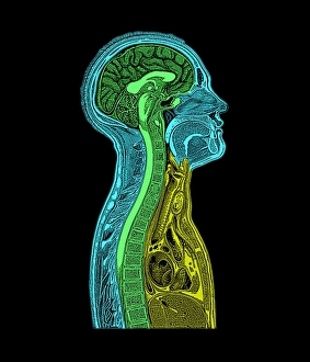
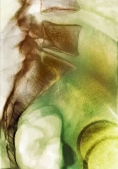
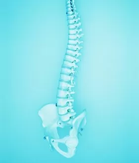
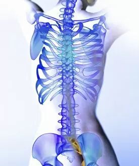
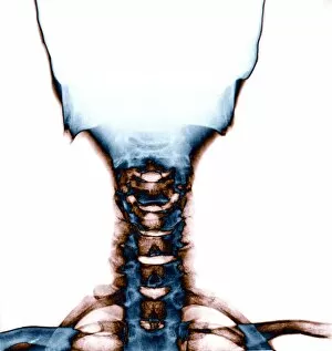
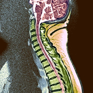
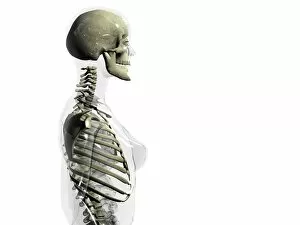
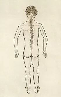
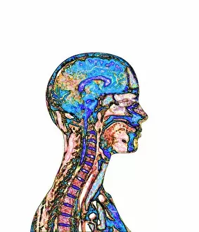
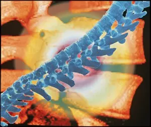
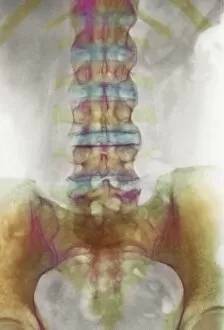
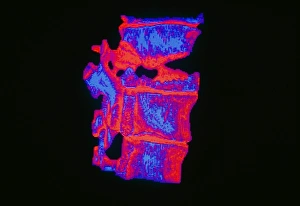
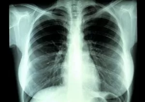
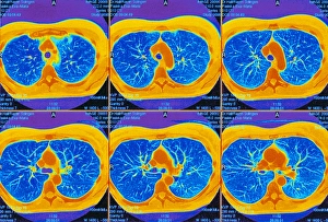
The backbone, also known as the spine, is a remarkable structure that serves as the foundation of our body's support system. Just like a diagram of the human brain and spinal column reveals the intricate connection between these vital components, our backbone plays a crucial role in maintaining our overall health. Similar to the DNA molecule that carries genetic information within every cell, our spine holds essential information about our physical well-being. It acts as a central hub for communication between different parts of the body through its network of nerves. Centuries ago, medical pioneers like Calots introduced groundbreaking techniques such as spinal surgery to address issues related to this complex structure. Their efforts paved the way for advancements in understanding and treating conditions affecting not only bones but also other aspects connected to it. Artwork depicting DNA molecules reminds us that even at a microscopic level, there is an inherent link between genetics and spine health. The neck and shoulder arteries visible on X-rays demonstrate how blood flow nourishes this critical area, ensuring optimal functioning. X-ray images showcasing normal spines give us insight into bone structure from a back view perspective. These visuals allow us to appreciate how each vertebra contributes to forming this sturdy yet flexible framework supporting our entire body. However, sometimes challenges arise such as slipped discs which can cause discomfort or pain. Artistic representations help illustrate these conditions visually while MRI scans provide detailed insights into their impact on normal torso structures. Ultimately, it is important not only to understand anatomy but also take care of our backbones by adopting healthy habits like regular exercise and proper posture. By appreciating both its complexity and resilience, we can ensure that this integral part of ourselves remains strong throughout life's journey.