
Axial View Collection
"Exploring the Intricacies of Axial View: Unveiling the Hidden Secrets within Medical Imaging" Delving into the Depths


All Professionally Made to Order for Quick Shipping
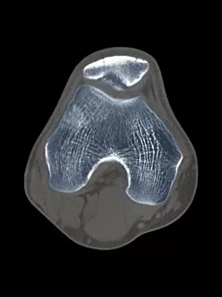
"Exploring the Intricacies of Axial View: Unveiling the Hidden Secrets within Medical Imaging" Delving into the Depths: A Glimpse of a Normal Brain through Axial MRI Scans Unraveling Spinal Mysteries: Axial View Reveals Herniated Disk, Compressed Spinal Cord, and Nerve Roots at C5 Peering into Precision: The Beauty of a Normal Axial View at C5 - Spotting Disks, Nerve Roots, and Posterior Structures Beyond Skin Deep: CT Scan F006/9140 Uncovers Knee Disease in Exquisite Detail Precision Matters: Targeted Breast Surgery Visualized Through Mammogram's Axial Perspective Ancient Clues Resurface: Homo heidelbergensis Vertebra C015/6799 Comes to Life with an Axial View Challenging Diagnosis Made Clearer: Exploring Abnormalities in a Cystic Pancreas Tumor via CT Scan's Axial Perspective Unmasking Lung Troubles: Identifying Lung Abscesses through Detailed CT Scans from an Axial Angle Revisiting Lung Abscesses' Enigma Once More - Insights from Additional CT Scans in the Same Plane Captivating Complexity.