Fraud Blocker Skip to main content

Arthropods Collection (page 4)

Arthropods: A Fascinating World of Color and Adaptation From the icy depths of Greenland to the vibrant gardens they can be found in every corner of our planet

Background imageArthropods Collection: Musca domestica and larva, fig. 399 from A. Celli, 'Manuale dell'igienista

Musca domestica and larva, fig. 399 from A. Celli, "Manuale dell'igienista, ad uso di ufficiali sanitari, medici circondariali e provinciali, ingegneri, chimici e veterinari igienisti

Background imageArthropods Collection: Glossina palpalis (Diptera. Brachycera. Muscidae), from A. Celli, 'Manuale dell'igienista

Glossina palpalis (Diptera. Brachycera. Muscidae), from A. Celli, "Manuale dell'igienista, ad uso di ufficiali sanitari, medici circondariali e provinciali, ingegneri

Background imageArthropods Collection: Culex pipiens larva (Culicidae), enlarged under the microscope

Culex pipiens larva (Culicidae), enlarged under the microscope. Date of Photograph:04/1914

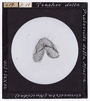
Background imageArthropods Collection: Tracheae of the proboscis of Musca domestica (Muscidae), enlarged under the microscope

Tracheae of the proboscis of Musca domestica (Muscidae), enlarged under the microscope. Date of Photograph:04/1894

Background imageArthropods Collection: Male Chironomus plumosus (Culicidiformes), magnified under the microscope

Male Chironomus plumosus (Culicidiformes), magnified under the microscope. Date of Photograph:06/12/1890

Background imageArthropods Collection: Head of male and female Culex pipiens and male and female Anopheles claviger

Head of male and female Culex pipiens and male and female Anopheles claviger, magnified under the microscope. Date of Photograph:1890-1914 ca

Background imageArthropods Collection: Male Culex pipiens (Culicidae), magnified under the microscope

Male Culex pipiens (Culicidae), magnified under the microscope. Date of Photograph:04/1895

Background imageArthropods Collection: Head of female Culex (Culicidae), enlarged under the microscope

Head of female Culex (Culicidae), enlarged under the microscope. Date of Photograph:06/12/1890

Background imageArthropods Collection: Head of male Anapheles (Diptera) enlarged under a microscope

Head of male Anapheles (Diptera) enlarged under a microscope. Date of Photograph:1872-1915 ca

Background imageArthropods Collection: Larva of Anopheles (Diptera) magnified under the microscope

Larva of Anopheles (Diptera) magnified under the microscope. Date of Photograph:1872-1915 ca

Background imageArthropods Collection: Stegomyia fasciata male and female. Yellow fever, fig. 398 from A. Celli, Manuale dell'igienista

Stegomyia fasciata male and female. Yellow fever, fig. 398 from A. Celli, Manuale dell'igienista, ad uso di ufficiali sanitari, medici circondariali e provinciali, ingegneri

Background imageArthropods Collection: Phthirius pubis (Pubic Lice) enlarged under a microscope

Phthirius pubis (Pubic Lice) enlarged under a microscope. Date of Photograph:04/1914

Background imageArthropods Collection: Microscopically enlarged sting of Vespa vulgaris (Vespidae)

Microscopically enlarged sting of Vespa vulgaris (Vespidae). Date of Photograph:04/1895

Background imageArthropods Collection: Musca domestica (Diptera. Muscidae), from A. Celli, 'Manuale dell'igienista

Musca domestica (Diptera. Muscidae), from A. Celli, "Manuale dell'igienista, ad uso di ufficiali sanitari, medici circondariali e provinciali, ingegneri, chimici e veterinari igienisti

Background imageArthropods Collection: Male Anopheles maculiformis, magnified under the microscope

Male Anopheles maculiformis, magnified under the microscope. Date of Photograph:1890-1914 ca

Background imageArthropods Collection: Pupa of Culex Pipiens magnified under a microscope

Pupa of Culex Pipiens magnified under a microscope. Date of Photograph:04/1914

Background imageArthropods Collection: Diaptomus Castor (Crostacei. Entomostracee) enlarged under a microscope

Diaptomus Castor (Crostacei. Entomostracee) enlarged under a microscope. Date of Photograph:11/1897

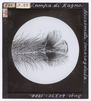
Background imageArthropods Collection: Micromata smaragdula spider leg, magnified under a microscope

Micromata smaragdula spider leg, magnified under a microscope. Date of Photograph:19/04/1894

Background imageArthropods Collection: Hyperia trigona. (Amphipoda. Hyperina) magnified under the microscope

Hyperia trigona. (Amphipoda. Hyperina) magnified under the microscope. Date of Photograph:04/1914

Background imageArthropods Collection: Gammarus pulex (Amphipoda, Gammaridae) magnified under the microscope

Gammarus pulex (Amphipoda, Gammaridae) magnified under the microscope. Date of Photograph:04/1914

Background imageArthropods Collection: Corophium longicorne (Amphipoda, Gammaridae) magnified under the microscope

Corophium longicorne (Amphipoda, Gammaridae) magnified under the microscope. Date of Photograph:04/1914

Background imageArthropods Collection: Pediculus Capitis (Apteri) enlarged under a microscope

Pediculus Capitis (Apteri) enlarged under a microscope. Date of Photograph:04/1914

Background imageArthropods Collection: Phylloxera Vastatrix root louse (Aphidae) enlarged under a microscope

Phylloxera Vastatrix root louse (Aphidae) enlarged under a microscope. Date of Photograph:1914

Background imageArthropods Collection: Cornea of the eye of a Tabanus Bovinus (Tabanidae) enlarged under a microscope

Cornea of the eye of a Tabanus Bovinus (Tabanidae) enlarged under a microscope. Date of Photograph:04/1895

Background imageArthropods Collection: Haematopota pluvialis (Diptera. Brachycera Fam. Tabanidae), from A. Celli, 'Manuale dell'igienista

Haematopota pluvialis (Diptera. Brachycera Fam. Tabanidae), from A. Celli, "Manuale dell'igienista, ad uso di ufficiali sanitari, medici circondariali e provinciali, ingegneri

Background imageArthropods Collection: Cordylobia anthropophaga female and larva (Diptera) from A. Celli, 'Manuale dell'igienista

Cordylobia anthropophaga female and larva (Diptera) from A. Celli, "Manuale dell'igienista, ad uso di ufficiali sanitari, medici circondariali e provinciali, ingegneri

Background imageArthropods Collection: Stomoxys calcitrans. (Diptera. Brachycera Muscidae), enlarged under the microscope from A

Stomoxys calcitrans. (Diptera. Brachycera Muscidae), enlarged under the microscope from A. Celli, "Manuale dell'igienista, ad uso di ufficiali sanitari, medici circondariali e provinciali

Background imageArthropods Collection: Psycoda phalaenoides (Culiciformes), enlarged under the microscope

Psycoda phalaenoides (Culiciformes), enlarged under the microscope. Date of Photograph:1914

Background imageArthropods Collection: Scatophaga leg fly, magnified under the microscope

Scatophaga leg fly, magnified under the microscope. Date of Photograph:19/04/1894

Background imageArthropods Collection: Head of male Anopheles, magnified under the microscope

Head of male Anopheles, magnified under the microscope. Date of Photograph:1900-1915 ca

Background imageArthropods Collection: Culex pipiens and Anopheles claviger larvae, enlarged under the microscope

Culex pipiens and Anopheles claviger larvae, enlarged under the microscope. Date of Photograph:1890-1914 ca

Background imageArthropods Collection: Head of female Anapheles (Diptera) enlarged under a microscope

Head of female Anapheles (Diptera) enlarged under a microscope. Date of Photograph:1872-1915 ca

Background imageArthropods Collection: Culex (Diptera) larvae enlarged under a microscope

Culex (Diptera) larvae enlarged under a microscope. Date of Photograph:1872-1915 ca

Background imageArthropods Collection: Culex pipiens and Anopheles maculipennis in resting state, fig. 22 from A

Culex pipiens and Anopheles maculipennis in resting state, fig. 22 from A. Celli, Manuale dell'igienista, ad uso di ufficiali sanitari, medici circondariali e provinciali, ingegneri

Background imageArthropods Collection: Culex and Anopheles eggs enlarged, fig. 19 from A. Celli, Manuale dell'igienista

Culex and Anopheles eggs enlarged, fig. 19 from A. Celli, Manuale dell'igienista, ad uso di ufficiali sanitari, medici circondariali e provinciali, ingegneri, chimici e veterinari igienisti

Background imageArthropods Collection: Dytiscus Natator (Dytiscidae) enlarged under a microscope

Dytiscus Natator (Dytiscidae) enlarged under a microscope. Date of Photograph:1914

Background imageArthropods Collection: Pulex canis (Pulicidae) magnified under the microscope

Pulex canis (Pulicidae) magnified under the microscope. Date of Photograph:15/04/1894

Background imageArthropods Collection: A dragonfly and a flying deer, pen drawing on reddish paper by Leonardo da Vinci

A dragonfly and a flying deer, pen drawing on reddish paper by Leonardo da Vinci and preserved at the Royal Library of Turin

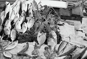
Background imageArthropods Collection: School of fish

School of fish Palermo. Date of Photograph:1960-1961

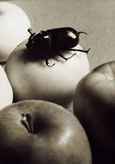
Background imageArthropods Collection: A coleoptera on an apple. Postcard sent by the photographer to Vincenzo Balocchi

A coleoptera on an apple. Postcard sent by the photographer to Vincenzo Balocchi Italy. Date of Photograph:1942

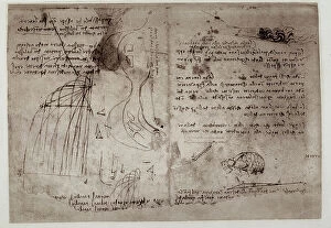
Background imageArthropods Collection: Study on waterfalls, written by Leonardo da Vinci, part of the Arundel Codex 263, c.36r

Study on waterfalls, written by Leonardo da Vinci, part of the Arundel Codex 263, c.36r, housed in the British Museum of London Drawing High Renaissance, Renaissance

Background imageArthropods Collection: Two crabs; pen drawing on white paper by Leonardo da Vinci preserved in the Wallraff-Richartz

Two crabs; pen drawing on white paper by Leonardo da Vinci preserved in the Wallraff-Richartz Museum in Cologne. Drawing High Renaissance, Renaissance, Renaissance-Baroque styles and periods, Europe

Background imageArthropods Collection: Still life with flowers, fruit and insects; painting by Rachel Ruysch

Still life with flowers, fruit and insects; painting by Rachel Ruysch. Galleria Palatina, Palazzo Pitti, Florence Painting Eighteenth Century, Renaissance-Baroque styles and periods, Europe

Background imageArthropods Collection: Still life with fruit and flowers; painting of Rachel Ruysch

Still life with fruit and flowers; painting of Rachel Ruysch. Galleria Palatina, Palazzo Pitti, Florence Painting Eighteenth Century, Renaissance-Baroque styles and periods, Europe

Background imageArthropods Collection: Table full of food

Table full of food France. Date of Photograph:1907-1910

Background imageArthropods Collection: Bas relief depicting a crab, allegory of the sign of the zodiac, Cancer

Bas relief depicting a crab, allegory of the sign of the zodiac, Cancer. Sacrament Chapel or Chapel of the Planets. Work by Agostino di Duccio

Background imageArthropods Collection: Bas relief depicting a scorpio, allegory of the sign of the zodiac, Scorpio

Bas relief depicting a scorpio, allegory of the sign of the zodiac, Scorpio. Sacrament Chapel or Chapel of the Planets. Work by Agostino di Duccio

Background imageArthropods Collection: Blazon of Pope Urbano VIII Barberini, in the Basilica of S. Pietro, Rome

Blazon of Pope Urbano VIII Barberini, in the Basilica of S. Pietro, Rome. Rome San Pietro Church Sculpture Baroque, Renaissance-Baroque styles and periods, Europe, First and Second Millennium A.D


All Professionally Made to Order for Quick Shipping

Arthropods: A Fascinating World of Color and Adaptation From the icy depths of Greenland to the vibrant gardens they can be found in every corner of our planet. Take a closer look at these incredible creatures and discover their captivating beauty. In the frigid waters, a massive Greenland shark gracefully glides through the darkness. Attached to its body is a parasitic copepod, Ommatokoita elongata, relying on this majestic predator for survival. Moving from water to land, bee anatomy reveals intricate structures designed for efficiency. Historical artwork showcases the meticulous details that have fascinated scientists and artists alike throughout centuries. Basking under the warm sun, a Red Admiral butterfly rests delicately on a plant. Its wings spread wide open, displaying an exquisite pattern that mesmerizes all who behold it. Another Red Admiral butterfly takes flight with elegance and grace. As its wings flutter effortlessly in mid-air, it becomes one with nature's symphony of colors. The sea green swallowtail butterfly dances among flowers with unmatched gracefulness. Its delicate movements mirror the gentle waves caressing sandy shores. A circle of bees buzzes harmoniously as they work together towards a common goal – building their hive and collecting nectar from blooming flowers. Exploring pond life reveals an enchanting world teeming with diverse arthropods. Mantis shrimp roam coral reefs in Puerto Galera while water fleas swim alongside green algae in garden ponds - showcasing nature's interconnectedness. A hornet mimic hoverfly cleverly disguises itself as one of nature's fiercest predators – evoking both fear and admiration for its remarkable adaptation skills. Amongst them all stands proudly the Peacock butterfly adorned with striking patterns resembling eyespots on its wings. Small tortoiseshell butterflies join this display of natural artistry - reminding us that even small creatures possess immense beauty worth cherishing and can truly extraordinary beings.