
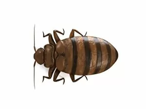
Animal Anatomy Collection (page 2)
"Exploring the Intricacies of Animal Anatomy: A Glimpse into the Past" Step back in time to 1832 and immerse yourself in the captivating world of animal anatomy


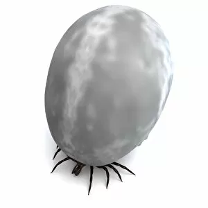
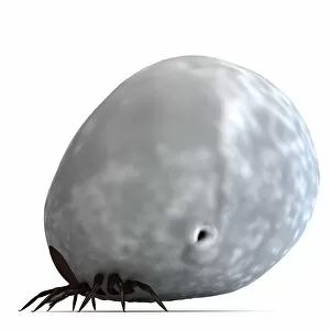


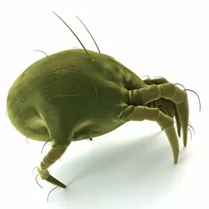


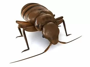


All Professionally Made to Order for Quick Shipping
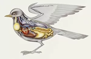
"Exploring the Intricacies of Animal Anatomy: A Glimpse into the Past" Step back in time to 1832 and immerse yourself in the captivating world of animal anatomy. Delve into a treasure trove of hand-colored engravings, showcasing the remarkable skeletal structure of various creatures. First, let's unravel the secrets hidden within a dog skeleton. With its intricate network of bones, this artwork F006/2267 unveils the foundation that supports our loyal companions' every movement. Moving on to aquatic wonders, we encounter "The Haddock, " "The Shad, " and "The Pike. " These exquisite engravings from A Treatise on Fish and Fish-ponds offer a glimpse into these species' inner workings. Marvel at their streamlined bodies and finely crafted fins that enable them to navigate through water with grace. Venturing further underwater, we stumble upon "The Cod Fish" and "The Barbel. " These illustrations provide insight into how these fish adapt to their unique environments. Observe their specialized features designed for survival amidst ocean depths or river currents. Next up is an enchanting depiction titled "Salmon Trout from Berwick on Tweed. " This artwork captures the elegance of this magnificent creature as it swims against powerful currents—a testament to nature's resilience. "The Whiting, " another stunning engraving from A Treatise on Fish and Fish-ponds, showcases yet another marvel of animal anatomy. Its slender body hints at its ability to swiftly maneuver through vast oceans in search of sustenance. Continuing our journey along rivers, we encounter "The Pearch from the River Rhine. " The intricately illustrated scales reveal not only beauty but also protection—armor against potential predators lurking beneath tranquil waters. Our exploration wouldn't be complete without acknowledging one of nature's most abundant species—the humble herring. As depicted in this hand-colored engraving, it highlights both simplicity and complexity—an embodiment of the delicate balance within ecosystems.