
Anatomical Drawing Collection
Anatomical drawing is an art form that beautifully captures the intricate details of various organisms and human anatomy


All Professionally Made to Order for Quick Shipping
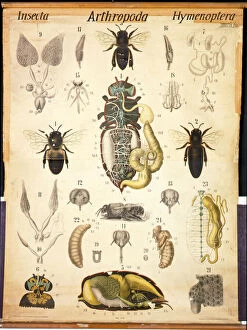
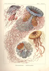
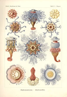
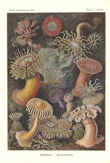
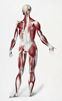
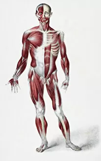
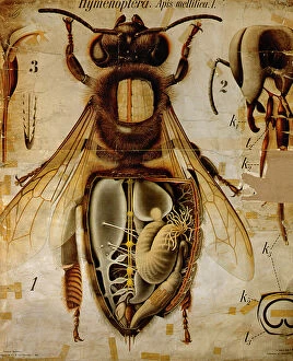
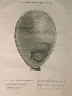
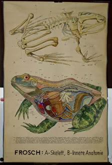
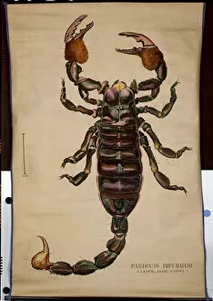
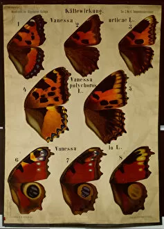
Anatomical drawing is an art form that beautifully captures the intricate details of various organisms and human anatomy. From the mesmerizing "Anatomy of the Apis" to the captivating "Discomedusae - Scheibenquallen, " these drawings provide a glimpse into the fascinating world of biology. One such masterpiece is "Acanthometra - Stachelstrahlinge, " which showcases the delicate beauty of these marine creatures. The vibrant colors and meticulous detailing make this artwork truly awe-inspiring. Similarly, "Asteridea - Sea Star" portrays the enchanting diversity found in our oceans, with each sea star exhibiting its unique characteristics. The mesmerizing depiction of jellyfish in "Siphonophorae" transports us to an ethereal underwater realm. The graceful movements captured in this artwork evoke a sense of wonder and curiosity about these mysterious creatures. Equally captivating is "Actiniae - Sea Anemone, " which showcases their stunning tentacles and vibrant hues, reminding us of nature's endless wonders. Moving from marine life to human anatomy, we encounter a surgical diagram from Johannes de Ketham's renowned work, Fasciculus Medicinae. This woodcut illustration provides invaluable insights into medical practices during that era, highlighting both scientific knowledge and artistic skill. Exploring further into human anatomy, we come across detailed drawings depicting muscles, sinews, and bones on both the front and back sides of male bodies. These illustrations offer a comprehensive understanding of our physical structure while showcasing the artist's ability to capture anatomical accuracy with precision. Lastly, let us not forget about other remarkable beings like honey bees whose intricate internal structures are revealed through Pfurtschellers Zoological Wall Chart's "Anatomy of Honey Bee. " This colorful lithograph allows us to appreciate their complex physiology while marveling at their vital role in nature. Anatomical drawings serve as windows into worlds unseen by the naked eye.