
Anatomical Collection
"Unveiling the Anatomical Wonders: Exploring the Intricacies of Life" Step into a world where science meets art, as we delve into the fascinating realm of anatomy


All Professionally Made to Order for Quick Shipping
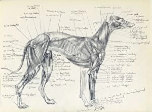
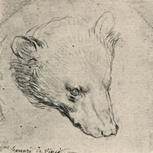
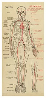
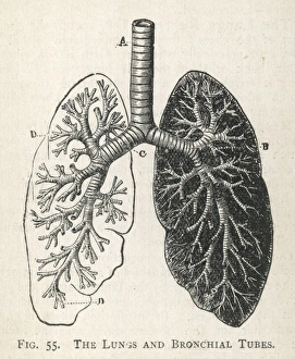
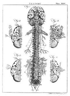
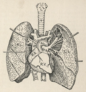
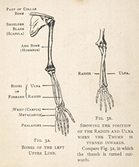
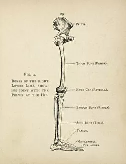
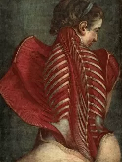
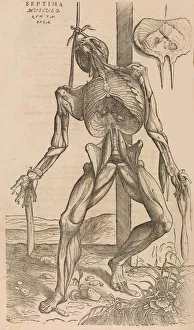
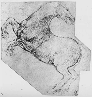
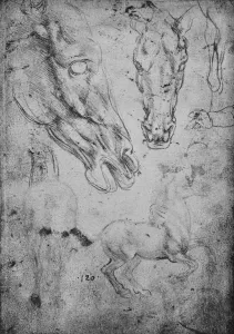
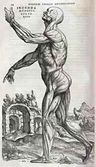
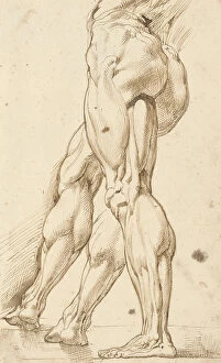
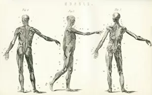
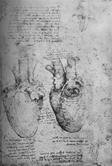
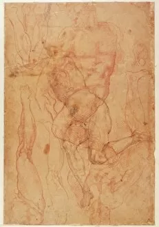
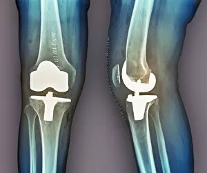
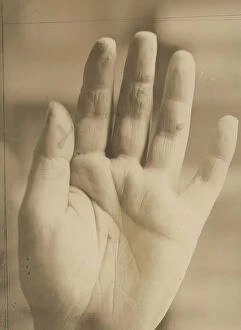
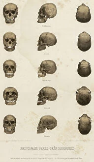
"Unveiling the Anatomical Wonders: Exploring the Intricacies of Life" Step into a world where science meets art, as we delve into the fascinating realm of anatomy. From Leonardo da Vinci's captivating depiction of a bear's head to his meticulous study of a greyhound's skeleton, these timeless masterpieces offer us glimpses into the intricate workings of living beings. As we journey further, we encounter detailed diagrams showcasing the complexity and beauty of our own bodies. The lungs and bronchial tubes reveal their delicate network, ensuring every breath sustains us. The human brain and spinal column stand as guardians of our thoughts and movements, orchestrating an orchestra within. Our exploration takes us deeper still, unveiling the heart's secrets through cross-sections that expose its innermost chambers. French wood engravings from the late 19th century transport us back in time to witness this vital organ in all its glory. But it is not just our internal structures that captivate; our limbs too hold stories waiting to be told. Diagrams meticulously illustrate each bone in hand and arm or right leg and hip – reminding us that beneath our skin lies an intricate framework supporting every step we take. And finally, a modern twist awaits with an MRI-style X-ray revealing a leg adorned with a stiletto shoe – merging technology with artistry while peering beyond appearances. From ancient manuscripts like "De humani corporis fabrica" to contemporary medical imaging techniques, these anatomical wonders remind us that beneath outward appearances lie marvels waiting to be discovered. They invite us to appreciate both the fragility and resilience encapsulated within each living being – truly celebrating life itself through understanding its intricacies.