Fraud Blocker Skip to main content

Alga Collection (page 6)

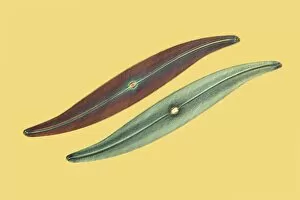
"Discovering the Intricate Beauty of Algae: From Ernst Haeckel's Art to 19th-Century Cyanotypes and SEM Images" Immerse yourself in the captivating world of algae

Background imageAlga Collection: LM of the red algae, Plumaria elegans C014 / 8488

LM of the red algae, Plumaria elegans C014 / 8488
Plumaria algae. Light micrograph of part of the finely branched thallus of the red algae Plumaria elegans (family: Florideophycidae)
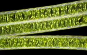
Background imageAlga Collection: LM of the red algae, Plumaria elegans C014 / 8487

LM of the red algae, Plumaria elegans C014 / 8487
Plumaria algae. Light micrograph of part of the finely branched thallus of the red algae Plumaria elegans (family: Florideophycidae)

Background imageAlga Collection: Diatom, light micrograph C016 / 8606

Diatom, light micrograph C016 / 8606
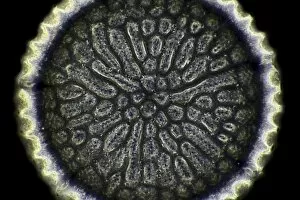
Diatom. Phase-contrast light micrograph of a diatom from the Mediterranean. Diatoms are a group of photosynthetic, single-celled algae containing about 10, 000 species

Background imageAlga Collection: Diatoms, light micrograph C016 / 8605

Diatoms, light micrograph C016 / 8605
Diatoms, Shearing contrast light micrograph. Diatoms are a group of photosynthetic, single-celled algae containing about 10, 000 species

Background imageAlga Collection: Diatoms, light micrograph C016 / 8598

Diatoms, light micrograph C016 / 8598
Diatoms. Interphase contrast light micrograph of a selection of differently shaped diatoms. Diatoms are a group of photosynthetic, single-celled algae containing about 10, 000 species

Background imageAlga Collection: Diatoms, light micrograph C016 / 8602

Diatoms, light micrograph C016 / 8602
Diatoms. Phase-contrast light micrograph of diatoms from the Mediterranean. Diatoms are a group of photosynthetic, single-celled algae containing about 10, 000 species

Background imageAlga Collection: Diatoms, light micrograph C016 / 8600

Diatoms, light micrograph C016 / 8600
Diatoms. Differential interference contrast (DIC) light micrograph of diatoms from the Mediterranean. Diatoms are a group of photosynthetic, single-celled algae containing about 10, 000 species

Background imageAlga Collection: Green algae, light micrograph C016 / 8595

Green algae, light micrograph C016 / 8595
Green algae, Interphase contrast light micrograph. Magnification: x280, when printed 10 centimetres wide

Background imageAlga Collection: Fossil diatom, light micrograph C016 / 8586

Fossil diatom, light micrograph C016 / 8586
Fossil diatom. Interphase contrast light micrograph of a fossilised diatom. Diatoms are a group of photosynthetic, single-celled algae containing about 10, 000 species

Background imageAlga Collection: Diatoms, light micrograph C016 / 8585

Diatoms, light micrograph C016 / 8585
Diatoms. Interphase contrast light micrograph of two differently shaped diatoms. Diatoms are a group of photosynthetic, single-celled algae containing about 10, 000 species

Background imageAlga Collection: Fossil diatom, light micrograph C016 / 8583

Fossil diatom, light micrograph C016 / 8583
Fossil diatom. Interphase contrast light micrograph of a fossilised diatom. Diatoms are a group of photosynthetic, single-celled algae containing about 10, 000 species

Background imageAlga Collection: Paramecium bursaria protozoan C016 / 8579

Paramecium bursaria protozoan C016 / 8579
Paramecium bursaria protozoan, phase-contrast light micrograph. This ciliate protozoan inhabits freshwater, where it feeds mainly on bacteria

Background imageAlga Collection: Plant-based insulating materials C014 / 0322

Plant-based insulating materials C014 / 0322
Plant-based insulating materials. Close-up of two different insulating materials made from plant products; one made from compressed dried algae (top) and the other made from soya (Glycine max)

Background imageAlga Collection: Microalgae research

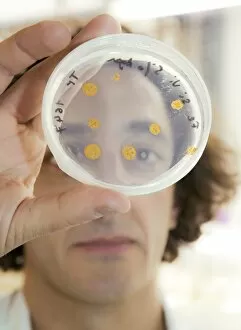
Microalgae research. Biologist holding a Petri dish containing cultured diatom microalgae. Diatoms are photosynthetic, single-celled algae that form an important part of the plankton at the base of

Background imageAlga Collection: Seaweed (Desmarestia ligulata) C016 / 6014

Seaweed (Desmarestia ligulata) C016 / 6014
seaweed (Desmarestia ligulata). Page 55 from Algae Danmonienses: or dried specimens of Marine Plants, principally collected in Devonshire by Mary Wyatt; carefully named according to Dr

Background imageAlga Collection: Algae-covered gravestone

Algae-covered gravestone. Orange-coloured filamentous green algae (Trentepohlia aurea) growing on a gravestone. Photographed in Culbone, Exmoor, Somerset, UK, in March

Background imageAlga Collection: Carrageen moss (Chondus cripus) C016 / 6062

Carrageen moss (Chondus cripus) C016 / 6062
Carrageen moss (Chondus cripus). Specimen from Algae Danmonienses: or dried specimens of Marine Plants, principally collected in Devonshire by Mary Wyatt; carefully named according to Dr

Background imageAlga Collection: Fossil-bearing rock, light micrograph C017 / 8482

Fossil-bearing rock, light micrograph C017 / 8482
Fossil-bearing rock, light micrograph. This thin slice from a flint rock includes fossils of algae and shells of gastropods (snails)

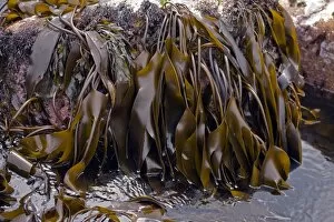
Background imageAlga Collection: Kelp seaweed C017 / 8437

Kelp seaweed C017 / 8437
Kelp seaweed (Laminaria digitata) on rocks at low tide. This seaweed is also known as devils apron and oarweed. Products such as ice cream, toothpaste, bread

Background imageAlga Collection: Coastal rock pool C017 / 8431

Coastal rock pool C017 / 8431
Coastal rock pool. Close-up of seaweed and other marine plants on the edge of a rock pool at low tide in a coastal area

Background imageAlga Collection: Coastal rock pool C017 / 8430

Coastal rock pool C017 / 8430
Coastal rock pool. Close-up of limpets and seaweed on the edge of a pool of seawater in rocks at low tide in a coastal area

Background imageAlga Collection: Coastal rocks and pebbles C017 / 8326

Coastal rocks and pebbles C017 / 8326
Coastal rocks and pebbles. These rocks and pebbles have formed by coastal erosion on the Lizard Peninsula, Cornwall, UK. The rocks and minerals include serpentine (black-green)

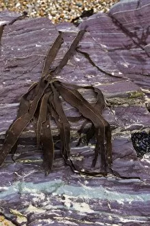
Background imageAlga Collection: Devonian slates C017 / 8323

Devonian slates C017 / 8323
Devonian slates and seaweed. Close-up of banded Devonian coastal rocks and coloured slates at Polzeath, Cornwall, UK. Slate is a metamorphic rock

Background imageAlga Collection: Paramecium bursaria protozoan, light micr

Paramecium bursaria protozoan, light micr
Paramecium bursaria protozoan, differential interference contrast light micrograph. This ciliate protozoan inhabits freshwater, where it feeds mainly on bacteria

Background imageAlga Collection: Blue green algae stigonema, LM C013 / 5286

Blue green algae stigonema, LM C013 / 5286
Light micrograph using differential interference contrast microscopy (DIC) of the filamentous blue-green algae (group name Cyanophycophyta), called stigonema

Background imageAlga Collection: LM of the red algae, Plumaria elegans C013 / 5279

LM of the red algae, Plumaria elegans C013 / 5279
Plumaria algae. False color light micrograph of part of the finely branched thallus of the red algae Plumaria elegans (family: Florideophycidae)

Background imageAlga Collection: Hamelin Pool Marine Nature Reserve

Hamelin Pool Marine Nature Reserve, Shark Bay, Western Australia. This area is famous for its stromatolites, mineralised microbial communities formed from cyanobacteria (blue-green algae)

Background imageAlga Collection: Pink salt, Australia

Pink salt, Australia
MODEL RELEASED. Pink salt. Woman holding pink salt that has evaporated out of a salt lake. The pink colour is caused by the microscopic algae Dunaliella salina, which produces a red pigment

Background imageAlga Collection: Pink salt lake, Australia

Pink salt lake, Australia
Pink salt lake. Pink salt that has evaporated out of a salt lake. The pink colour is caused by the microscopic algae Dunaliella salina, which produces a red pigment

Background imageAlga Collection: Pink lake, Australia

Pink lake, Australia
Pink lake. The colour of this salt lake is caused by the microscopic algae Dunaliella salina, which produces a red pigment. Photographed near Port Gregory, Western Australia

Background imageAlga Collection: Diploneis diatom, SEM C013 / 5110

Diploneis diatom, SEM C013 / 5110
Diploneis diatom, coloured scanning electron micrograph (SEM). Diatoms are single-celled photosynthetic algae, of which there are about 100, 000 species

Background imageAlga Collection: Coccolithophorid plankton, SEM C013 / 5109

Coccolithophorid plankton, SEM C013 / 5109
Coccolithophorid plankton. Coloured scanning electron micrograph (SEM) of the external mineralised structure (coccosphere) of a small marine algal organism called a coccolithophore

Background imageAlga Collection: LM of the colonial diatom Fragillaria sp

LM of the colonial diatom Fragillaria sp
Light micrograph of the diatom Fragillaria sp.. The filaments seen here are simple colonies of Fragillaria. They consist of a stack of diatoms which have failed to separate following cell division

Background imageAlga Collection: Colonial alga, Dinobryon

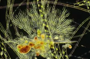
Colonial alga, Dinobryon
Light micrograph of a branching colony of the golden- brown alga Dinobryon sp.. The colony is composed of single-celled algae, each of which secretes a fine, transparent

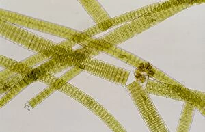
Background imageAlga Collection: Melosira filamentous diatom alage, LM

Melosira filamentous diatom alage, LM
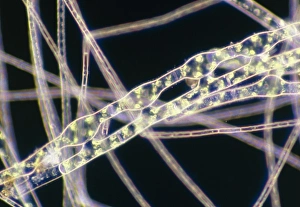
Melosira sp. diatoms. Light micrograph of filamentous strands of the freshwater diatom alga Melosira sp.. Diatoms are single-celled algae that form cell walls made of silica

Background imageAlga Collection: SEM of blue-green algae

SEM of blue-green algae
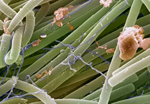
Blue-green algae. Coloured scanning electron micrograph (SEM) of Oscillatoria sp. filamentous blue-green algae. Blue-green algae are considered to be one of the most primitive organisms

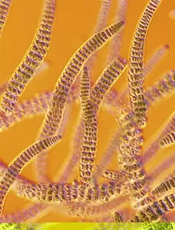
Background imageAlga Collection: Oscillatoria blue-green alga

Oscillatoria blue-green alga
Light micrograph of Oscillatoria sp. (brown strands), a filamentous blue-green alga or cyanobacterium. The strands of alga are in fact linear colonies of primitive, single-celled

Background imageAlga Collection: Diatom algae, Campylodiscus

Diatom algae, Campylodiscus
Diatoms. Coloured scanning electron micrograph of the diatom Campylodiscus hibernicus. The diatoms are a distinctive group of single-celled algae containing about 10, 000 species

Background imageAlga Collection: Colonial freshwater diatoms

Colonial freshwater diatoms
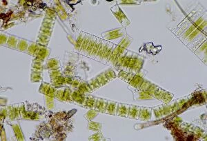
Light micrograph of assorted freshwater diatoms. The rectangular, broken filaments seen here are simple colonies. They consist of a chain of diatoms which have failed to separate following cell

Background imageAlga Collection: Light micrograph of assorted diatoms

Light micrograph of assorted diatoms. The diatoms are a distinctive group of single-celled algae containing about 10, 000 species

Background imageAlga Collection: Chlamydomonas sp. algae, SEM

Chlamydomonas sp. algae, SEM
Green algae. Coloured scanning electron micrograph (SEM) of two Chlamydomonas sp. green algae. These single-celled organisms are aquatic, living in freshwater habitats

Background imageAlga Collection: Green alga, Micrasterias

Green alga, Micrasterias
Micrasterias alga. Coloured scanning electron micrograph (SEM) of the desmid Micrasterias sp.. The desmids are a group of freshwater unicellular green algae characterised by their intricately- shaped

Background imageAlga Collection: Conjugating spirogyra

Conjugating spirogyra
Light micrograph showing conjugating filaments of Spirogyra, a green alga which forms slimy threads in stagnant or slow moving water

Background imageAlga Collection: LM of Chlorella surrounded by bacteria

LM of Chlorella surrounded by bacteria
Light micrograph of Chlorella sp. a unicellular green alga which inhabits freshwater ponds and lakes. The non-motile, globular cells may be solitary or aggregated into groups as seen here

Background imageAlga Collection: LM of conjugating filaments of Spirogyra

LM of conjugating filaments of Spirogyra
Light micrograph showing conjugating filaments of Spirogyra sp. a green alga which forms slimy threads in stagnant or slow moving water

Background imageAlga Collection: Filaments of Spirogyra alga

Filaments of Spirogyra alga
Light micrograph of filaments of Spirogyra, a green alga which forms slimy threads called water- silk" or " mermaids tresses" in stagnant or slow moving water

Background imageAlga Collection: Crab's Eye Lichen - UK

Crab's Eye Lichen - UK
DAC-820 Crab's Eye Lichen UK Ochrolechia parella David Chapman Please note that prints are for personal display purposes only and may not be reproduced in any way

Background imageAlga Collection: Scyphosphaera apsteinii

Scyphosphaera apsteinii. SEM image of an equatorial coccolith


All Professionally Made to Order for Quick Shipping

"Discovering the Intricate Beauty of Algae: From Ernst Haeckel's Art to 19th-Century Cyanotypes and SEM Images" Immerse yourself in the captivating world of algae, as we delve into its mesmerizing artistry and scientific wonders. Ernst Haeckel, a renowned biologist and artist, showcases the delicate intricacy of diatom algae through his stunning illustrations. These masterpieces reveal the symmetrical beauty hidden within these microscopic organisms. Step back in time with pressed seaweed specimens C016 / 6127 from centuries ago. Preserved meticulously, they offer a glimpse into the diverse forms and colors that algae can take on. The cyanotype technique used during the 19th century captures their essence in ethereal blue hues, adding an enchanting touch to their already fascinating nature. Calcareous phytoplankton observed under scanning electron microscopy (SEM) unravels another layer of algal marvels. Discosphaera tubifera, a coccolithophore species adorned with intricate calcium carbonate plates, showcases nature's architectural genius at work. Dictyota dichotoma emerges as a striking example of brown algae thriving along coastlines worldwide. Its branching structure creates an artistic spectacle underwater while providing essential habitats for marine life. Diatoms continue to amaze us with their geometric patterns when viewed under SEM imaging techniques. Their microscopic beauty is unveiled through meticulous detailing that reveals their remarkable diversity and ecological significance. Venturing further into aquatic realms, Fucus bulbosus or kelp stands tall as one of nature's giants beneath ocean waves. This majestic alga plays a crucial role in coastal ecosystems by offering shelter to numerous marine organisms. Pond life surprises us with its vibrant assortment of microorganisms where diatoms once again steal the show under SEM imaging techniques. Their intricate designs resemble miniature works of art floating amidst water bodies' tranquility.