Fraud Blocker Skip to main content

Abdominal Collection (page 3)

"Unleash the Power of Your Abdominal Strength: From Ancient Artwork to Modern Fitness" Athletic young man in shorts with a ball lying on a stone floor

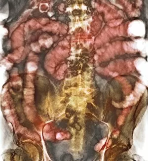
Background imageAbdominal Collection: Small bowel obstruction, X-ray C014 / 1818

Small bowel obstruction, X-ray C014 / 1818
Small bowel obstruction. Coloured X-ray of the abdomen of a 77 year old male patient with a small bowel obstruction and gallstones (white outlined spheres at top left)

Background imageAbdominal Collection: Trunk muscle anatomy, 1831 artwork

Trunk muscle anatomy, 1831 artwork
Trunk muscle anatomy. Coronal section of the trunk (anterior view) showing the anatomy of its muscles and bones. This anatomical artwork is plate 75 from volume 2 (1831)

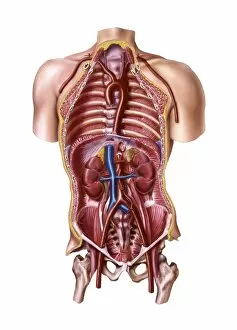
Background imageAbdominal Collection: Human blood vessel anatomy, artwork

Human blood vessel anatomy, artwork
Human blood vessel anatomy. Cutaway computer artwork showing the location of the major arteries and veins inside the human trunk. The kidneys (red, centre left and right) are also shown

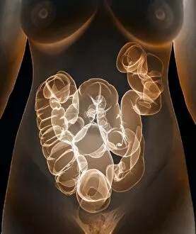
Background imageAbdominal Collection: Healthy large intestine, 3D CT scan C016 / 6509

Healthy large intestine, 3D CT scan C016 / 6509
Healthy large intestine. Coloured 3D computed tomography (CT) scan of the abdomen of a 37 year old female patient showing a healthy large intestine

Background imageAbdominal Collection: Abdominal pain C016 / 9665

Abdominal pain C016 / 9665
MODEL RELEASED. Abdominal pain

Background imageAbdominal Collection: Abdominal aorta, 3D CT scan C016 / 6542

Abdominal aorta, 3D CT scan C016 / 6542
Abdominal aorta. Coloured 3D computed tomography (CT) scan of the abdominal aorta of a 56 year old patient. The aorta is the bodys main artery. It carries blood from the heart (at top) around the body

Background imageAbdominal Collection: Intestinal hernia, artwork C016 / 7525

Intestinal hernia, artwork C016 / 7525
Intestinal hernia. Computer artwork of a person with an intestinal hernia (lower right), showing how it has been treated (left)

Background imageAbdominal Collection: Healthy large intestine, 3D CT scan C016 / 6508

Healthy large intestine, 3D CT scan C016 / 6508
Healthy large intestine. Coloured 3D computed tomography (CT) scan of the abdomen of a 37 year old female patient showing a healthy large intestine

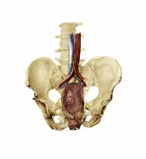
Background imageAbdominal Collection: Total mesorectal excision, artwork C016 / 6996

Total mesorectal excision, artwork C016 / 6996
Total mesorectal excision (TME). Artwork of the pelvis and the area of the colon and rectum that is removed in total mesorectal excision

Background imageAbdominal Collection: Healthy large intestine, 3D CT scan C016 / 6507

Healthy large intestine, 3D CT scan C016 / 6507
Healthy large intestine. Coloured 3D computed tomography (CT) scan of the abdomen of a 37 year old female patient showing a healthy large intestine

Background imageAbdominal Collection: Abdominal pain C016 / 9666

Abdominal pain C016 / 9666
MODEL RELEASED. Abdominal pain

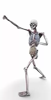
Background imageAbdominal Collection: Male skeleton and organs, artwork

Male skeleton and organs, artwork
Male skeleton and organs and blood vessels, artwork

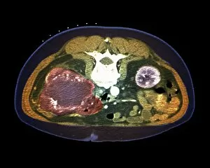
Background imageAbdominal Collection: Kidney tumour, CT scan

Kidney tumour, CT scan
Kidney tumour. Coloured computed tomography (CT) scan of a section through the abdomen of a 62-year-old patient, showing necrosis (death) of tissue in the right kidney

Background imageAbdominal Collection: Healthy abdomen, MRI scan C018 / 0663

Healthy abdomen, MRI scan C018 / 0663
Healthy abdomen. Coloured magnetic resonance imaging (MRI) scan of the abdomen of a 35 year old patient showing the internal organs. At top are the stomach (purple) and liver (red)

Background imageAbdominal Collection: Healthy abdomen, MRI scan C018 / 0664

Healthy abdomen, MRI scan C018 / 0664
Healthy abdomen. Coloured magnetic resonance imaging (MRI) scan of the abdomen of a 35 year old patient showing the internal organs. At top are the stomach (brown) and liver (orange)

Background imageAbdominal Collection: Ant anus, SEM C018 / 0549

Ant anus, SEM C018 / 0549
Ant anus, coloured scanning electron micrograph (SEM). Ants excrete bodily waste through a single orifice, their anus, which is located at the tip of the abdomen

Background imageAbdominal Collection: Abdominal pain and headache C014 / 1285

Abdominal pain and headache C014 / 1285
MODEL RELEASED. Abdominal pain and headache. Woman holding her painful abdomen and head. Common causes of abdominal pain include food poisoning, indigestion, menstrual cramps, and strained muscles

Background imageAbdominal Collection: Abdominal pain C014 / 1283

Abdominal pain C014 / 1283
MODEL RELEASED. Abdominal pain. Woman holding her painful abdomen. Common causes of abdominal pain include food poisoning, indigestion, menstrual cramps, and strained muscles

Background imageAbdominal Collection: Abdominal pain C014 / 1284

Abdominal pain C014 / 1284
MODEL RELEASED. Abdominal pain. Woman holding her painful abdomen. Common causes of abdominal pain include food poisoning, indigestion, menstrual cramps, and strained muscles

Background imageAbdominal Collection: Small bowel obstruction, X-ray C014 / 1819

Small bowel obstruction, X-ray C014 / 1819
Small bowel obstruction. Coloured X-ray of the abdomen of a 77 year old male patient with a small bowel obstruction and gallstones (spheres at top left). The white areas are gas-filled bowel loops

Background imageAbdominal Collection: Rectal polyp removal, artwork

Rectal polyp removal, artwork
Rectal polyp removal. Artwork showing the use of a colonoscope to remove a polyp or cyst from the inside of the rectum. The rectum (down left) is the final part of the digestive system

Background imageAbdominal Collection: Ovarian cyst in abdomen, artwork

Ovarian cyst in abdomen, artwork
Ovarian cyst in abdomen. Artwork of a large ovarian cyst (centre left) in a dissected human abdomen, seen from above. The uterus (centre) and the ovaries are part of the female reproductive system

Background imageAbdominal Collection: Abdominal muscles, artwork

Abdominal muscles, artwork
Abdominal muscles. Computer artwork of the rectus abdominis muscles (centre) and the transverse abdominis muscles (right and left)

Background imageAbdominal Collection: Female groin arteries, 1825 artwork

Female groin arteries, 1825 artwork
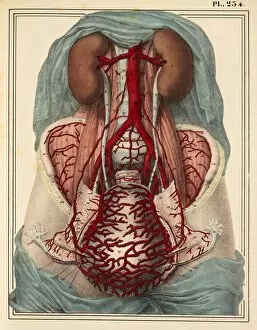
Female groin arteries. Dissections of a female groin to show areas supplied by the internal iliac artery (red) and its branches

Background imageAbdominal Collection: Lumbar spinal nerves, 1825 artwork

Lumbar spinal nerves, 1825 artwork
Lumbar spinal nerves. Lateral view of the lower vertebral column, showing nerves (white) of the sympathetic nervous system. The pelvic ilium bone has been removed

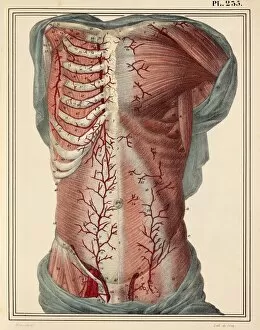
Background imageAbdominal Collection: Trunk arteries, 1825 artwork

Trunk arteries, 1825 artwork
Trunk arteries. Dissection showing the internal mammary arteries (red, in chest) and the epigastric arteries (red, in groin and abdomen), from a mans trunk (torso)

Background imageAbdominal Collection: Lateral trunk muscles, 1831 artwork

Lateral trunk muscles, 1831 artwork
Lateral trunk muscles. Side view (front of body at right) of the third layer of the lateral muscles of the trunk. The muscles include the serratus, the transverse abdominal

Background imageAbdominal Collection: Liver and stomach arteries, 1825 artwork

Liver and stomach arteries, 1825 artwork
Liver and stomach arteries. Dissection of the abdomen, showing the arteries (red) of the liver (brown, upper left), stomach (pink, upper centre), and the intestines (grey with yellow fat)

Background imageAbdominal Collection: Veins of the stomach, 1825 artwork

Veins of the stomach, 1825 artwork
Veins of the stomach. Abdominal dissection showing the veins (blue) of the stomach (centre) and the greater omentum (lower centre, yellow)

Background imageAbdominal Collection: Trunk musculature, 1831 artwork

Trunk musculature, 1831 artwork
Trunk musculature, anterior view. This anatomical artwork is plate 62 from volume 2 (1831) of Traite complet de l anatomie de l homme (1831-1854)

Background imageAbdominal Collection: Intestinal lymph vessels, 1825 artwork

Intestinal lymph vessels, 1825 artwork
Intestinal lymph vessels. Dissection of the abdominal cavity showing lymphatic vessels (white) and nodes (lumps). The thoracic duct (top) connects with the intestinal lymph trunk (centre)

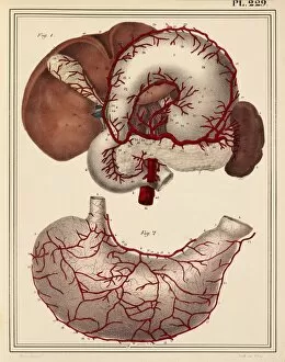
Background imageAbdominal Collection: Digestive organ arteries, 1825 artwork

Digestive organ arteries, 1825 artwork
Digestive organ arteries. Arteries (red) of various organs of the digestive system. At bottom are arteries of the posterior side of the stomach

Background imageAbdominal Collection: Uterine arteries, 1825 artwork

Uterine arteries, 1825 artwork
Uterine arteries. Dissection showing the arteries (red) of the uterus (lower centre) of a woman who died six days after giving birth. The ovaries and fallopian tubes are either side

Background imageAbdominal Collection: Groin muscles, 1831 artwork

Groin muscles, 1831 artwork
Groin muscles. Muscles of the anterior wall of the lower (groin) region of the trunk, with a study of the inguinal canal. This anatomical artwork is plate 70 from volume 2 (1831)

Background imageAbdominal Collection: Internal body organs, 1825 artwork

Internal body organs, 1825 artwork
Internal body organs. Dissection showing the internal organs of the chest and abdomen. The overlying tissues have been removed to show (from top) the lungs

Background imageAbdominal Collection: Human pancreas, artwork

Human pancreas, artwork
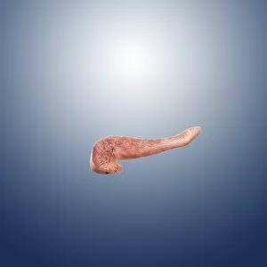
Human pancreas, computer artwork. The pancreas secretes digestive enzymes as well as hormones, including insulin

Background imageAbdominal Collection: Human anatomy, 1823 C017 / 8057

Human anatomy, 1823 C017 / 8057
Human anatomy. 19th-century illustrations showing the anatomy of a human skull (upper left), brain and spinal column (upper centre), chest and abdominal organs (lungs, heart, liver, stomach)

Background imageAbdominal Collection: Urinary system, artwork C015 / 6455

Urinary system, artwork C015 / 6455
Urinary system. Computer artwork showing the organs of the human urinary system. The kidneys (red, top left and right) filter waste products such as urea from the blood (red and blue) to form urine

Background imageAbdominal Collection: Urinary system, artwork C015 / 6454

Urinary system, artwork C015 / 6454
Urinary system. Computer artwork showing the organs of the human urinary system. The kidneys (red, top left and right) filter waste products such as urea from the blood to form urine

Background imageAbdominal Collection: Large bowel obstruction, X-ray C017 / 7855

Large bowel obstruction, X-ray C017 / 7855
Large bowel obstruction. X-ray of the abdomen of a patient with an obstructed large intestine. The obstruction may be due to cancer, twisting of the intestine, an injury or an infection

Background imageAbdominal Collection: Large bowel obstruction, X-ray C017 / 7854

Large bowel obstruction, X-ray C017 / 7854
Large bowel obstruction. X-ray of the abdomen of a patient with an obstructed large intestine. The obstruction may be due to cancer, twisting of the intestine, an injury or an infection


All Professionally Made to Order for Quick Shipping

"Unleash the Power of Your Abdominal Strength: From Ancient Artwork to Modern Fitness" Athletic young man in shorts with a ball lying on a stone floor: This dedicated athlete knows that strong abdominal muscles are key to his performance and agility. Cardiovascular system meets historical artwork: Explore how the ancient Greeks depicted the importance of a well-toned abdomen in their sculptures, recognizing its connection to overall health and vitality. Young man in a suit and without shirt lying on a couch: Even amidst busy schedules, this gentleman understands the significance of maintaining core strength for good posture and stability. Young man in a suit and without shirt kneeling on a couch: Witness the dedication as this individual engages his abdominal muscles while working from home, demonstrating that fitness can be integrated into any routine. Male groin arteries, 1825 artwork: Delve into medical illustrations from centuries ago, showcasing early anatomical studies highlighting the intricate network of blood vessels within our abdominal region. Man with naked torso wearing long underwear in horse barn sitting on bales of hay: Embrace unconventional workout spaces as we witness this rugged individual utilizing his surroundings to engage his core muscles effectively. Woman using an exercise ball: Discover how incorporating an exercise ball into your fitness routine can target your abs while improving balance and flexibility for optimal results. Healthy lower back X-ray reveals strong abdominals' impact: Peek inside at an X-ray image showcasing how well-developed abdominal muscles provide support for our spine, preventing lower back pain or injuries. Abdominal organs - The hidden heroes within us all. Gain insight into these vital organs residing within our abdomen that play crucial roles in digestion, metabolism, immunity, and more. Sir Astley Paston Cooper's legacy lives on through understanding abdominals' importance - Learn about Sir Astley Paston Cooper's pioneering work in surgical anatomy, including his contributions to understanding the abdominal region.