Home > Arts > Artists > S > George Stubbs
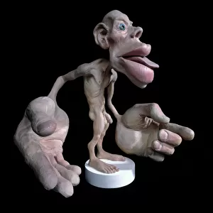
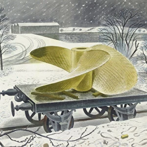
Study of the Human Figure, Posterior View, from A Comparative Anatomical Exposition of the Structure of the Human Body with that of a Tiger and a Common Fowl, c.1795-1806 (graphite & ink on paper)
")
![]()
Wall Art and Photo Gifts from Fine Art Finder
Study of the Human Figure, Posterior View, from A Comparative Anatomical Exposition of the Structure of the Human Body with that of a Tiger and a Common Fowl, c.1795-1806 (graphite & ink on paper)
XYC257414 Study of the Human Figure, Posterior View, from A Comparative Anatomical Exposition of the Structure of the Human Body with that of a Tiger and a Common Fowl, c.1795-1806 (graphite & ink on paper) by Stubbs, George (1724-1806); 48.9x33 cm; Yale Center for British Art, Paul Mellon Collection, USA
Media ID 33068768
© Bridgeman Images
Anatomist Biology Bottom Diagram Dorsal Feet George Stubbs Internal Anatomy Legs Limbs Muscular Proportion Rear Recto Stubbs George 1724 1806 Veins Britisch Britisch
FEATURES IN THESE COLLECTIONS
> Animals
> Mammals
> Cats (Wild)
> Tiger
> Arts
> Artists
> S
> George Stubbs
> Arts
> Realistic drawings
> Figure drawing
> Fine art portraits
> Arts
> Realistic drawings
> Graphite art
> Fine art
> Fine Art Finder
> Artists
> George Stubbs
> Fine Art Finder
> Science,scientists & Inventions
> Sport
> Sports Stars
> Paul George
EDITORS COMMENTS
This print showcases George Stubbs' "Study of the Human Figure, Posterior View" from his renowned work "A Comparative Anatomical Exposition of the Structure of the Human Body with that of a Tiger and a Common Fowl" created between 1795-1806. The artwork, measuring 48.9x33 cm, is held in the prestigious Paul Mellon Collection at the Yale Center for British Art in the USA. Stubbs, an eminent English artist and anatomist (1724-1806), meticulously depicted the internal anatomy of both humans and animals throughout his career. In this particular study, he focuses on capturing the posterior view of a human figure with remarkable precision using graphite and ink on paper. The piece not only reflects Stubbs' exceptional artistic skills but also highlights his deep understanding of anatomy. His ability to merge art and science seamlessly is evident as he portrays intricate details such as muscles, bones, and organs. This print serves as a testament to Stubbs' contribution to medical illustration during the late 18th century. It stands as a valuable resource for anatomists and medical professionals alike who seek to understand human physiology more comprehensively. Bernie C. Staggers skillfully captures this significant artwork through photography, allowing viewers to appreciate its beauty even from afar. Through this image reproduction by Fine Art Finder, we can continue to admire Stubbs' genius while acknowledging Staggers' talent in preserving artistic masterpieces for generations to come.
MADE IN THE UK
Safe Shipping with 30 Day Money Back Guarantee
FREE PERSONALISATION*
We are proud to offer a range of customisation features including Personalised Captions, Color Filters and Picture Zoom Tools
SECURE PAYMENTS
We happily accept a wide range of payment options so you can pay for the things you need in the way that is most convenient for you
* Options may vary by product and licensing agreement. Zoomed Pictures can be adjusted in the Basket.