Fraud Blocker Skip to main content

Home > All Images > 2013 > April > 2 Apr 2013

Images Dated 2nd April 2013 (page 3)

272 items

Background imageImages Dated 2nd April 2013: The Martin Luther King Memorial on the National Mall in Washington, D. C

The Martin Luther King Memorial on the National Mall in Washington, D. C. United States of America, North America
The Martin Luther King Memorial on the National Mall in Washington, D.C. United States of America, North America

Background imageImages Dated 2nd April 2013: The Lincoln Memorial, Washington, D. C. United States of America, North America

The Lincoln Memorial, Washington, D. C. United States of America, North America
The Lincoln Memorial, Washington, D.C. United States of America, North America

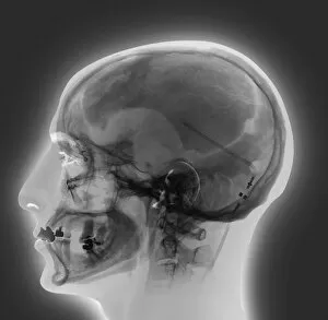
Background imageImages Dated 2nd April 2013: Ventricular shunt, X-ray

Ventricular shunt, X-ray
Ventricular shunt. Coloured X-ray of the head of a 52-year-old patient with hydrocephalus, showing the position of a ventricular shunt (light lines, right) and its external valve (round)

Background imageImages Dated 2nd April 2013: Dodder seed, SEM C015 / 5077

Dodder seed, SEM C015 / 5077
Dodder (Cuscuta sp.) sede, coloured environmental scanning electron micrograph (ESEM)

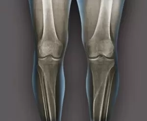
Background imageImages Dated 2nd April 2013: Normal knees, X-rays

Normal knees, X-rays
Normal knees. Coloured frontal X-rays of the healthy knees of a 35-year-old

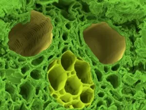
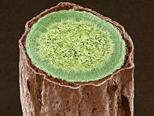
Background imageImages Dated 2nd April 2013: Bamboo stem, SEM C015 / 5073

Bamboo stem, SEM C015 / 5073
Bamboo stem. Coloured environmental scanning electron micrograph (ESEM) of a section through the stem of a bamboo plant (family Gramineae), showing a vascular bundle

Background imageImages Dated 2nd April 2013: Houttuynia cordata leaf surface, SEM C015 / 5078

Houttuynia cordata leaf surface, SEM C015 / 5078
Leaf surface. Coloured environmental scanning electron micrograph (ESEM) of two trichomes (hair-like) on a Houttuynia cordata leaf. These are glandular trichomes, which produce an orange-scented oil

Background imageImages Dated 2nd April 2013: Ventricular shunt, X-ray

Ventricular shunt, X-ray
Ventricular shunt. Coloured X-ray of the head of a 38-year-old patient with hydrocephalus, showing the position of a ventricular shunt (dark lines) and its external valve (round, centre right)

Background imageImages Dated 2nd April 2013: Ventricular shunt, X-ray

Ventricular shunt, X-ray
Ventricular shunt. Coloured X-ray of the head of a 38-year-old patient with hydrocephalus, showing the position of a ventricular shunt (dark lines) and its external valve (round, centre right)

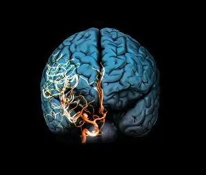
Background imageImages Dated 2nd April 2013: Brain aneurysm, 3D scan

Brain aneurysm, 3D scan
Brain aneurysm. 3D computed tomography (CT) angiogram coupled with a magnetic resonance imaging (MRI) scan of the brain of a 38-year-old, showing a large aneurysm (bright)

Background imageImages Dated 2nd April 2013: Coronary stenosis before treatment, X-ray

Coronary stenosis before treatment, X-ray
Coronary stenosis before treatment. Coloured angiogram of the left coronary artery in the heart of a 60-year-old patient, showing severe narrowing (stenosis)

Background imageImages Dated 2nd April 2013: Coronary stenosis treatment, X-ray

Coronary stenosis treatment, X-ray
Coronary stenosis treatment. Coloured angiogram of a balloon catheter (white tube, centre right) inserted into the anterior interventricular branch of the left coronary artery in the heart of a

Background imageImages Dated 2nd April 2013: Coronary stenosis before treatment, X-ray

Coronary stenosis before treatment, X-ray
Coronary stenosis before treatment. Angiogram of the left coronary artery in the heart of a 60-year-old patient, showing severe narrowing (stenosis)

Background imageImages Dated 2nd April 2013: Ventricular shunt, X-ray

Ventricular shunt, X-ray
Ventricular shunt. Coloured X-ray of the head of a 52-year-old patient with hydrocephalus, showing the position of a ventricular shunt (light lines, right) and its external valve (round)

Background imageImages Dated 2nd April 2013: Brain aneurysm, 3D scan

Brain aneurysm, 3D scan
Brain aneurysm. 3D computed tomography (CT) angiogram coupled with a magnetic resonance imaging (MRI) scan of the brain of a 38-year-old, showing a large aneurysm (bright)

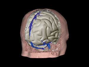
Background imageImages Dated 2nd April 2013: Thrombophlebitis in the brain, 3D CT scan

Thrombophlebitis in the brain, 3D CT scan
Thrombophlebitis in the brain. Coloured 3D computed tomography (CT) angiogram of the blood vessels (blue) in the brain of a 48-year-old with thrombophlebitis of the superior sagittal sinus

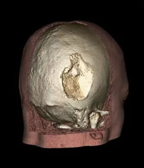
Background imageImages Dated 2nd April 2013: Gorhams disease, 3D CT scan

Gorhams disease, 3D CT scan
Gorhams disease. Coloured 3D computed tomography (CT) scan of the rear of the skull of a 47-year-old patient, showing damage (centre) caused by osteolysis (bone resorption and replacement)

Background imageImages Dated 2nd April 2013: Gorhams disease, 3D CT scan

Gorhams disease, 3D CT scan
Gorhams disease. Coloured 3D computed tomography (CT) scan of the rear of the skull of a 47-year-old patient, showing damage (centre) caused by osteolysis (bone resorption and replacement)

Background imageImages Dated 2nd April 2013: Lichen, SEM C015 / 5082

Lichen, SEM C015 / 5082
Lichen, coloured environmental scanning electron micrograph (ESEM). Lichens are symbiotic organisms formed of a fungus and an alga that mutually benefit one another

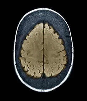
Background imageImages Dated 2nd April 2013: Thickened skull, MRI scan

Thickened skull, MRI scan
Thickened skull. Coloured magnetic resonance imaging (MRI) scan of an axial section through the head of a 22-year-old patient, showing thickening of the trabecular (spongy) bone of the skull

Background imageImages Dated 2nd April 2013: Coronary stenosis treatment, X-ray

Coronary stenosis treatment, X-ray
Coronary stenosis treatment. Angiogram of a balloon catheter (white tube, centre right) inserted into the anterior interventricular branch of the left coronary artery in the heart of a 60-year-old

Background imageImages Dated 2nd April 2013: Coronary stenosis after treatment, X-ray

Coronary stenosis after treatment, X-ray
Coronary stenosis after treatment. Angiogram of the left coronary artery in the heart of a 60-year-old patient after balloon catheterisation

Background imageImages Dated 2nd April 2013: Gramicidin antibiotic molecule C015 / 5071

Gramicidin antibiotic molecule C015 / 5071
Gramicidin. Molecular model of the antibiotic gramicidin D, from the bacterium Bacillus brevis. This antibiotic is effective against Gram positive and Gram negative bacteria

Background imageImages Dated 2nd April 2013: Paralympics crowds

Paralympics crowds. Photographed on 8 September 2012, at the Olympic Stadium during the London 2012 Paralympics

Background imageImages Dated 2nd April 2013: Ventricular shunt, X-ray

Ventricular shunt, X-ray
Ventricular shunt. X-ray of the head of a 52-year-old patient with hydrocephalus, showing the position of a ventricular shunt (dark lines, right) and its external valve (round, centre right)

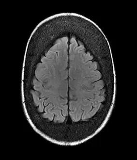
Background imageImages Dated 2nd April 2013: Thickened skull, MRI scan

Thickened skull, MRI scan
Thickened skull. Magnetic resonance imaging (MRI) scan of an axial section through the head of a 22-year-old patient, showing thickening of the trabecular (spongy) bone of the skull

Background imageImages Dated 2nd April 2013: Brain aneurysm, 3D scan

Brain aneurysm, 3D scan
Brain aneurysm. 3D computed tomography (CT) angiogram coupled with a magnetic resonance imaging (MRI) scan of the brain of a 38-year-old, showing a large aneurysm (bright)

Background imageImages Dated 2nd April 2013: Thickened skull, MRI scan

Thickened skull, MRI scan
Thickened skull. Coloured magnetic resonance imaging (MRI) scan of a coronal section through the head of a 22-year-old patient, showing thickening of the trabecular (spongy) bone of the skull

Background imageImages Dated 2nd April 2013: Ribonuclease bound to angiogenin C015 / 5069

Ribonuclease bound to angiogenin C015 / 5069
Ribonuclease bound to angiogenin. Molecular model of a molecule of human angiogenin bound to a human placental ribonuclease inhibitor molecule

Background imageImages Dated 2nd April 2013: Gorhams disease, CT scans

Gorhams disease, CT scans
Gorhams disease. Coloured 3D (left) and 2D (right) computed tomography (CT) scans of the skull of a 47-year-old patient, showing damage (centre) caused by osteolysis (bone resorption and replacement)

Background imageImages Dated 2nd April 2013: Ventricular shunt, X-ray

Ventricular shunt, X-ray
Ventricular shunt. Coloured X-ray of the head of a 52-year-old patient with hydrocephalus, showing the position of a ventricular shunt (dark lines, right) and its external valve (round)

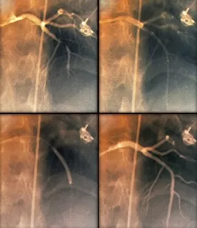
Background imageImages Dated 2nd April 2013: Coronary stenosis treatment, X-rays

Coronary stenosis treatment, X-rays
Coronary stenosis treatment. Coloured angiograms showing the left coronary artery in the heart of a 60-year-old patient before (top left)

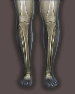
Background imageImages Dated 2nd April 2013: Normal legs, X-rays

Normal legs, X-rays
Normal legs. Coloured frontal X-rays of the healthy legs, of a 35-year-old

Background imageImages Dated 2nd April 2013: Coronary stenosis after treatment, X-ray

Coronary stenosis after treatment, X-ray
Coronary stenosis after treatment. Coloured angiogram of the left coronary artery in the heart of a 60-year-old patient after balloon catheterisation

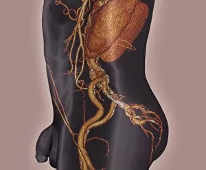
Background imageImages Dated 2nd April 2013: Atherosclerosis, 3D CT scan

Atherosclerosis, 3D CT scan
Atherosclerosis. 3D computed tomography (CT) angiogram of the main blood vessels of the lower body a 58-year-old man, showing numerous atheromatous plaques (light, centre) on the vessel walls

Background imageImages Dated 2nd April 2013: Paralympics track race

Paralympics track race. Photographed on 8 September 2012, at the Olympic Stadium during the London 2012 Paralympics

Background imageImages Dated 2nd April 2013: Thrombophlebitis in the brain, 3D CT scan

Thrombophlebitis in the brain, 3D CT scan
Thrombophlebitis in the brain. Coloured 3D computed tomography (CT) angiogram of the blood vessels (blue) in the brain of a 48-year-old with thrombophlebitis of the superior sagittal sinus

Background imageImages Dated 2nd April 2013: Winter on Exmoor

Winter on Exmoor. Winter sun shining on a wooded and snowy landscape on Exmoor. This area is in Somerset, UK

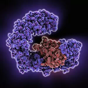
Background imageImages Dated 2nd April 2013: Importin protein complexed with enzyme C015 / 5149

Importin protein complexed with enzyme C015 / 5149
Importin protein complexed with enzyme, molecular model. Human importin13 (purple) complexed with SUMO-conjugating enzyme Ubc9 (red)

Background imageImages Dated 2nd April 2013: Importin protein complexed with enzyme C015 / 5148

Importin protein complexed with enzyme C015 / 5148
Importin protein complexed with enzyme, molecular model. Human importin13 (green) complexed with SUMO-conjugating enzyme Ubc9 (pink)

Background imageImages Dated 2nd April 2013: Divinity School, University of Oxford

Divinity School, University of Oxford, UK. This building, part of the Old Bodleian Library, is one of the oldest surviving buildings of the University of Oxford

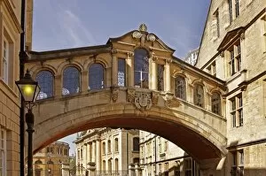
Background imageImages Dated 2nd April 2013: Bridge of Sighs, Oxford

Bridge of Sighs, Oxford. This skyway, the Hertford Bridge, crosses New College Lane and connects two buildings of Hertford College, part of the University of Oxford, UK

Background imageImages Dated 2nd April 2013: Passion flower hybrid cultivar

Passion flower hybrid cultivar (Passiflora edulis x caerulea Excel )

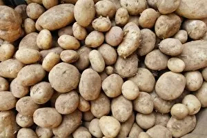
Background imageImages Dated 2nd April 2013: Organic potatoes

Organic potatoes (Solanum tuberosum). Potatoes are a good source of carbohydrate, providing energy in the diet. Organic farming reduces the use of pesticides and other synthetic chemicals

Background imageImages Dated 2nd April 2013: Velib rental bicycles, Paris

Velib rental bicycles, Paris
Velib rental bicycles. Row of Velib bicycles for hire on a street in Paris, France. These bikes are hired on a subscription and deposit basis

Background imageImages Dated 2nd April 2013: Magnolia stem, SEM C015 / 5128

Magnolia stem, SEM C015 / 5128
Magnolia stem. Coloured environmental scanning electron micrograph (ESEM) of a transverse section through the stem of a Magnolia sp. plant

Background imageImages Dated 2nd April 2013: Magnolia stem, SEM C015 / 5124

Magnolia stem, SEM C015 / 5124
Magnolia stem. Coloured environmental scanning electron micrograph (ESEM) of a transverse section through the stem of a Magnolia sp. plant

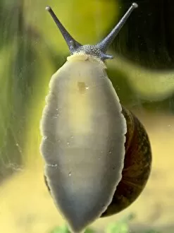
Background imageImages Dated 2nd April 2013: Garden snail climbing on glass

Garden snail climbing on glass
Garden snail (Helix aspersa) climbing on glass. The snails foot (the underside gripping area) and mouth are pressed to the surface of the glass


All Professionally Made to Order for Quick Shipping


-

Why Choose Us?

We are a leading provider of Art Prints and Photo Gifts since 2003, working in partnership with a range of Sporting Clubs, Charities, Museums and Picture Libraries. A large share of profits from any of their images will go directly towards supporting that charity or club. Our archive of images is carefully curated to bring you a wide range of subjects, including landscapes, wildlife, architecture, and more. We ship from our partner labs in the UK, USA, EU (Netherlands) and Australia.
+

How do I place an order?

Ordering is quick & easy - Just follow 5 Simple Steps:
  1. Find your image: Use our search box or browse our online photo Collections to find the image you want.

  2. Choose your print format: Select your desired product and add it to your cart.

  3. Enter your details: If you're a returning customer, simply enter your email address and password, and we'll fill in your billing and shipping address details. All personal details are held securely and are fully GDPR compliant. As standard, we remove all Personally Identifiable Information after 12 months.

  4. Pay for your purchase: We use state-of-the-art security for online shopping and do not have access to your card details.

  5. Sit back and relax: We'll email you confirmation of your order and when it's dispatched. Registered customers can also track orders in the 'My Account' area.

+

How do I pay for an order?

You can pay for your order with most of the major credit and debit cards, or PayPal. For added security, major financial institutions process payment details separately and securely on our behalf. We do not have access to your online payment card details. Online payments are preferred however we do take phone orders. For UK customers only we accept cheques issued against a UK bank.
+

Is my data safe?

We take data security very seriously. We do not have access to your full card details and all payments are requested over a fully secure connection. Additionally, we fully comply with current European and GDPR legislation, and automatically remove all personal data after at most 12 months (unless you have an account with us and have signed in recently). We also have a strict opt-in policy and would never sell your personal details. Your data is only used to fulfill your order promptly and efficiently.
+

Is wall art ready to hang?

For quick and easy installation all wall art, including framed prints, canvas prints and metal prints are supplied with a ready to hang solution on the back. Generally, saw tooth hangers are applied as they allow wall art to hang flush against the wall. The serrated edge of the hanger prevents the frame from shifting or tilting when hung.
+

Are the photo prints fade resistant?

Yes, we use archival quality photo paper photographic paper for vivid reproduction Prints are an accurate representations of the original artwork, which is preserved for artistic character and authenticity. We guarantee they match previews shown on our web site