Fraud Blocker Skip to main content

Home > All Images > 2012 > March > 7 Mar 2012

Images Dated 7th March 2012 (page 6)

338 items

Background imageImages Dated 7th March 2012: Marvin Hinton - Charlton Athletic

Marvin Hinton - Charlton Athletic
Football - 1961 / 1962 League Cup - First Round: Oldham Athletic 1 Charlton Athletic 4 Charltons Marvin Hinton at Boundary Park

Background imageImages Dated 7th March 2012: Tarantula hair, SEM

Tarantula hair, SEM
Tarantula hair. Coloured scanning electron micrograph (SEM) of a hair from the abdomen of a tarantula (family Theraphosidae) spider

Background imageImages Dated 7th March 2012: Fat cell, TEM

Fat cell, TEM
Fat cell. Coloured Coloured transmission electron micrograph (TEM) of a section through fat (adipose) tissue. Fat cells, or adipocytes

Background imageImages Dated 7th March 2012: Lymphocyte white blood cell, TEM

Lymphocyte white blood cell, TEM
Lymphocyte white blood cell. Coloured transmission electron micrograph (TEM) of a section through a lymphocyte from a lymph node

Background imageImages Dated 7th March 2012: Millipede head, SEM

Millipede head, SEM
Millipede head. Coloured scanning electron micrograph (SEM) of the head of a millipede from Barbados. Millipedes (class Diplopoda) have elongated bodies with two pairs of legs on each body segment

Background imageImages Dated 7th March 2012: Firefly head, SEM

Firefly head, SEM
Firefly head. Coloured scanning electron micrograph (SEM) of the head of a firefly (family Lampyridae), showing one of its large compound eyes (grey)

Background imageImages Dated 7th March 2012: 3D crystals, stereo artwork

3D crystals, stereo artwork. This image is designed to be viewed through 3D glasses

Background imageImages Dated 7th March 2012: Spinal cord, SEM

Spinal cord, SEM
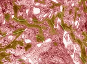
Spinal cord. Coloured scanning electron micrograph (SEM) of a section through a spinal cord, showing the grey matter (green) and white matter (blue)

Background imageImages Dated 7th March 2012: Lymph node, TEM

Lymph node, TEM
Lymph node. Coloured transmission electron micrograph (TEM) of a section through the peripheral corona of a lymph nodule, showing numerous small lymphocyte white blood cells (dark)

Background imageImages Dated 7th March 2012: Cell nucleus, TEM

Cell nucleus, TEM
Cell nucleus. Coloured transmission electron micrograph (TEM) of a section through a cell, showing the nucleus (large, spherical), and mitochondria (green)

Background imageImages Dated 7th March 2012: Fat cells, TEM

Fat cells, TEM
Fat cells. Coloured Coloured transmission electron micrograph (TEM) of a section through fat (adipose) tissue. Each fat cell, or adipocyte

Background imageImages Dated 7th March 2012: Millipede, SEM

Millipede, SEM
Millipede. Coloured scanning electron micrograph (SEM) of a millipede from Barbados. Millipedes (class Diplopoda) have elongated bodies with two pairs of legs on each body segment

Background imageImages Dated 7th March 2012: Glomerular capillary, TEM

Glomerular capillary, TEM
Glomerular capillary. Coloured transmission electron micrograph (TEM) of a section through kidney tissue, showing a capillary from a glomerulus. Glomeruli filter waste products from the blood

Background imageImages Dated 7th March 2012: Fat cell, TEM

Fat cell, TEM
Fat cell. Coloured Coloured transmission electron micrograph (TEM) of a section through fat (adipose) tissue. Fat cells, or adipocytes

Background imageImages Dated 7th March 2012: Macrophage white blood cell, TEM

Macrophage white blood cell, TEM
Macrophage white blood cell. Coloured transmission electron micrograph (TEM) of a section through a macrophage from a lymph node, showing the nucleus (red) and numerous lysosomes (black)

Background imageImages Dated 7th March 2012: Middle ear bone, SEM

Middle ear bone, SEM
Middle ear bone. Coloured scanning electron micrograph (SEM) of the human middle ear, showing the stapes (ring-shaped). The stapes is one of three bones (known as the ossicles)

Background imageImages Dated 7th March 2012: Higgs boson, artwork

Higgs boson, artwork
Higgs boson. Artwork representing the elementary particle known as the Higgs boson (centre). The existence of this hypothetical particle is predicted by the Standard Model of particle physics

Background imageImages Dated 7th March 2012: Retina blood vessel, SEM

Retina blood vessel, SEM
Retina blood vessel. Coloured scanning electron micrograph (SEM) of a section through a blood vessel in the retina of an eye, showing the red blood cells (red)

Background imageImages Dated 7th March 2012: Gram-negative bacterium, artwork

Gram-negative bacterium, artwork
Gram-negative bacterium. Artwork of a prokaryotic, Gram-negative rod-shaped bacterium with a single polar flagellum (spiral structure, upper left)

Background imageImages Dated 7th March 2012: Cell nucleus, TEM

Cell nucleus, TEM
Cell nucleus. Coloured transmission electron micrograph (TEM) of a section through a cell, showing the uncles (large, spherical), and mitochondria (blue)

Background imageImages Dated 7th March 2012: Particle physics experiment, artwork

Particle physics experiment, artwork
Particle physics experiment. Artwork of tracks of particles detected following a collision in a particle accelerator. In these experiments

Background imageImages Dated 7th March 2012: Eardrum, SEM

Eardrum, SEM
Eardrum. Coloured scanning electron micrograph (SEM) of an eardrum (red). The eardrum, or tympanic membrane, is located in the middle ear

Background imageImages Dated 7th March 2012: Glomerular capillary, TEM

Glomerular capillary, TEM
Glomerular capillary. Coloured transmission electron micrograph (TEM) of a section through kidney tissue, showing a capillary from a glomerulus. Glomeruli filter waste products from the blood

Background imageImages Dated 7th March 2012: Iris pigment epithelium, SEM

Iris pigment epithelium, SEM
Iris pigment epithelium. Coloured scanning electron micrograph (SEM) of a section through the iris of an eye, showing the iris pigment epithelium (IPE)

Background imageImages Dated 7th March 2012: Skin cell desmosomes, TEM

Skin cell desmosomes, TEM
Skin cell desmosomes. Coloured transmission electron micrograph (TEM) of a section through skin cells, showing numerous desmosomes (dark areas)

Background imageImages Dated 7th March 2012: Booklouse, SEM

Booklouse, SEM
Booklouse. Coloured scanning electron micrograph (SEM) of the head of a booklouse (order Psocoptera). These insects, also known as barklice or barkflies

Background imageImages Dated 7th March 2012: Cholera toxin, artwork

Cholera toxin, artwork
Cholera toxin, molecular structure. Cholera is an infectious intestinal disease caused by this toxin produced by the Gram-negative bacterium Vibrio cholerae

Background imageImages Dated 7th March 2012: Arteriole, TEM

Arteriole, TEM
Arteriole. Coloured transmission electron micrograph (TEM) of a section through the wall of an arteriole, showing smooth muscle (brown), endothelial cells (green)

Background imageImages Dated 7th March 2012: Nuclear research, conceptual artwork

Nuclear research, conceptual artwork
Nuclear research. Conceptual artwork of a scientist observing an atomic explosion in a petri dish. This image can represent research into nuclear weapons

Background imageImages Dated 7th March 2012: Firefly, SEM

Firefly, SEM
Firefly. Coloured scanning electron micrograph (SEM) of a firefly (family Lampyridae). These winged beetles, also known as lightning bugs, are named for their ability to flash their abdomens

Background imageImages Dated 7th March 2012: Firefly head, SEM

Firefly head, SEM
Firefly head. Coloured scanning electron micrograph (SEM) of the head of a firefly (family Lampyridae), showing one of its large compound eyes (grey)

Background imageImages Dated 7th March 2012: Fat cells, TEM

Fat cells, TEM
Fat cells. Coloured Coloured transmission electron micrograph (TEM) of a section through fat (adipose) tissue. Each fat cell, or adipocyte

Background imageImages Dated 7th March 2012: Millipede head, SEM

Millipede head, SEM
Millipede head. Coloured scanning electron micrograph (SEM) of the head of a millipede from Barbados. Millipedes (class Diplopoda) have elongated bodies with two pairs of legs on each body segment

Background imageImages Dated 7th March 2012: Froghopper, SEM

Froghopper, SEM
Froghopper. Coloured scanning electron micrograph (SEM) of the underside of the head of a froghopper (superfamily Cercopoidea)

Background imageImages Dated 7th March 2012: Firefly, SEM

Firefly, SEM
Firefly. Coloured scanning electron micrograph (SEM) of a firefly (family Lampyridae). These winged beetles, also known as lightning bugs, are named for their ability to flash their abdomens

Background imageImages Dated 7th March 2012: Optic nerve fibres, SEM

Optic nerve fibres, SEM
Optic nerve fibres. Coloured scanning electron micrograph (SEM) of axons from the optic nerve. Connective tissue fibres (thread-like) are also visible

Background imageImages Dated 7th March 2012: Spinal cord, SEM

Spinal cord, SEM
Spinal cord. Coloured scanning electron micrograph (SEM) of a section through a spinal cord, showing the grey matter (centre, pink) and white matter (red)

Background imageImages Dated 7th March 2012: Early universe, artwork

Early universe, artwork
Early universe. Artwork showing the clumping of matter in stellar nurseries in the early universe. Stars form when gravitational accretion causes clumps of gas and dust (nebulae)

Background imageImages Dated 7th March 2012: Brain lining, SEM

Brain lining, SEM
Brain lining. Coloured scanning electron micrograph (SEM) of the lining of the brain, showing the ciliary hairs (green) of ependymal cells

Background imageImages Dated 7th March 2012: Tarantula hair, SEM

Tarantula hair, SEM
Tarantula hair. Coloured scanning electron micrograph (SEM) of a hair from the abdomen of a tarantula (family Theraphosidae) spider

Background imageImages Dated 7th March 2012: Iris pigment epithelium, SEM

Iris pigment epithelium, SEM
Iris pigment epithelium. Coloured scanning electron micrograph (SEM) of a section through the iris of an eye, showing the iris pigment epithelium (IPE)

Background imageImages Dated 7th March 2012: Freshwater shrimp, SEM

Freshwater shrimp, SEM
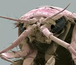
Freshwater shrimp. Coloured scanning electron micrograph (SEM) of the head of a freshwater shrimp (Gammarus pulex), showing its large eyes (black)

Background imageImages Dated 7th March 2012: Millipede, SEM

Millipede, SEM
Millipede. Coloured scanning electron micrograph (SEM) of a millipede from Barbados. Millipedes (class Diplopoda) have elongated bodies with two pairs of legs on each body segment

Background imageImages Dated 7th March 2012: Macrophage white blood cell, TEM

Macrophage white blood cell, TEM
Macrophage white blood cell. Coloured transmission electron micrograph (TEM) of a section through a macrophage from a lymph node, showing the nucleus (red) and numerous lysosomes (black)

Background imageImages Dated 7th March 2012: Middle ear bone, SEM

Middle ear bone, SEM
Middle ear bone. Coloured scanning electron micrograph (SEM) of the human middle ear, showing the stapes (ring-shaped). The stapes is one of three bones (known as the ossicles)

Background imageImages Dated 7th March 2012: Lymphocyte white blood cell, TEM

Lymphocyte white blood cell, TEM
Lymphocyte white blood cell. Coloured transmission electron micrograph (TEM) of a section through a lymphocyte from a lymph node

Background imageImages Dated 7th March 2012: Freshwater shrimp, SEM

Freshwater shrimp, SEM
Freshwater shrimp. Coloured scanning electron micrograph (SEM) of the head of a freshwater shrimp (Gammarus pulex), showing its large eyes (dark red)

Background imageImages Dated 7th March 2012: Lymph node, TEM

Lymph node, TEM
Lymph node. Coloured transmission electron micrograph (TEM) of a section through the peripheral corona of a lymph nodule, showing numerous small lymphocyte white blood cells (dark)


All Professionally Made to Order for Quick Shipping


-

Why Choose Us?

We are a leading provider of Art Prints and Photo Gifts since 2003, working in partnership with a range of Sporting Clubs, Charities, Museums and Picture Libraries. A large share of profits from any of their images will go directly towards supporting that charity or club. Our archive of images is carefully curated to bring you a wide range of subjects, including landscapes, wildlife, architecture, and more. We ship from our partner labs in the UK, USA, EU (Netherlands) and Australia.
+

How do I place an order?

Ordering is quick & easy - Just follow 5 Simple Steps:
  1. Find your image: Use our search box or browse our online photo Collections to find the image you want.

  2. Choose your print format: Select your desired product and add it to your cart.

  3. Enter your details: If you're a returning customer, simply enter your email address and password, and we'll fill in your billing and shipping address details. All personal details are held securely and are fully GDPR compliant. As standard, we remove all Personally Identifiable Information after 12 months.

  4. Pay for your purchase: We use state-of-the-art security for online shopping and do not have access to your card details.

  5. Sit back and relax: We'll email you confirmation of your order and when it's dispatched. Registered customers can also track orders in the 'My Account' area.

+

How do I pay for an order?

You can pay for your order with most of the major credit and debit cards, or PayPal. For added security, major financial institutions process payment details separately and securely on our behalf. We do not have access to your online payment card details. Online payments are preferred however we do take phone orders. For UK customers only we accept cheques issued against a UK bank.
+

Is my data safe?

We take data security very seriously. We do not have access to your full card details and all payments are requested over a fully secure connection. Additionally, we fully comply with current European and GDPR legislation, and automatically remove all personal data after at most 12 months (unless you have an account with us and have signed in recently). We also have a strict opt-in policy and would never sell your personal details. Your data is only used to fulfill your order promptly and efficiently.
+

Is wall art ready to hang?

For quick and easy installation all wall art, including framed prints, canvas prints and metal prints are supplied with a ready to hang solution on the back. Generally, saw tooth hangers are applied as they allow wall art to hang flush against the wall. The serrated edge of the hanger prevents the frame from shifting or tilting when hung.
+

Are the photo prints fade resistant?

Yes, we use archival quality photo paper photographic paper for vivid reproduction Prints are an accurate representations of the original artwork, which is preserved for artistic character and authenticity. We guarantee they match previews shown on our web site