Fraud Blocker Skip to main content

Home > All Images > 2011 > October > 21 Oct 2011

Images Dated 21st October 2011 (page 9)

830 items

Background imageImages Dated 21st October 2011: Cross section biomedical illustration of large and small intestine and pelvis in adult female

Cross section biomedical illustration of large and small intestine and pelvis in adult female

Background imageImages Dated 21st October 2011: Cross section biomedical illustration of esophagus, liver, gallbladder, pancreas in adult female

Cross section biomedical illustration of esophagus, liver, gallbladder, pancreas in adult female

Background imageImages Dated 21st October 2011: Cross section biomedical illustration of liver lobule

Cross section biomedical illustration of liver lobule

Background imageImages Dated 21st October 2011: Cross section biomedical illustration of colonoscopy procedure in adult female

Cross section biomedical illustration of colonoscopy procedure in adult female

Background imageImages Dated 21st October 2011: Cross section biomedical illustration of hernia repair and site of incision to uncover the hernia

Cross section biomedical illustration of hernia repair and site of incision to uncover the hernia

Background imageImages Dated 21st October 2011: Biomedical illustration of umbilical hernia in female baby

Biomedical illustration of umbilical hernia in female baby

Background imageImages Dated 21st October 2011: Cross section biomedical illustration of position of colectomy incision

Cross section biomedical illustration of position of colectomy incision

Background imageImages Dated 21st October 2011: Cross section biomedical illustration of human cell division with zygote dividing to form new cells

Cross section biomedical illustration of human cell division with zygote dividing to form new cells

Background imageImages Dated 21st October 2011: Cross section biomedical illustration of mitosis where the nucleus breaks down

Cross section biomedical illustration of mitosis where the nucleus breaks down and threads form across cell with chromosomes lining up on these threads

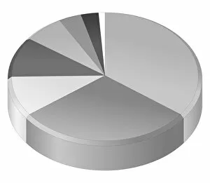
Background imageImages Dated 21st October 2011: Illustration of pie chart

Illustration of pie chart

Background imageImages Dated 21st October 2011: Cross section biomedical illustration of tumour-forming cancer cell

Cross section biomedical illustration of tumour-forming cancer cell

Background imageImages Dated 21st October 2011: Cross section biomedical illustration of cancer cells forming tumour at first doubling stage

Cross section biomedical illustration of cancer cells forming tumour at first doubling stage

Background imageImages Dated 21st October 2011: Cross section biomedical illustration of cancer cells forming tumour at second doubling stage

Cross section biomedical illustration of cancer cells forming tumour at second doubling stage

Background imageImages Dated 21st October 2011: Cross section biomedical illustration of cancer cells forming tumour at third doubling stage

Cross section biomedical illustration of cancer cells forming tumour at third doubling stage

Background imageImages Dated 21st October 2011: Cross section biomedical illustration of cancer cells forming tumour at fourth doubling stage

Cross section biomedical illustration of cancer cells forming tumour at fourth doubling stage

Background imageImages Dated 21st October 2011: Cross section biomedical illustration of Angiogenesis - how tumour obtains nutrients

Cross section biomedical illustration of Angiogenesis - how tumour obtains nutrients

Background imageImages Dated 21st October 2011: Cross section biomedical illustration of digestive tract in adult female

Cross section biomedical illustration of digestive tract in adult female

Background imageImages Dated 21st October 2011: Cross section biomedical illustration of damaged skin and blood vessel

Cross section biomedical illustration of damaged skin and blood vessel

Background imageImages Dated 21st October 2011: Cross section biomedical illustration of blood clotting over broken skin and fibroblasts multiplying

Cross section biomedical illustration of blood clotting over broken skin and fibroblasts multiplying and migrating to injury

Background imageImages Dated 21st October 2011: Cross section biomedical illustration of adult female human skeletal system and joints

Cross section biomedical illustration of adult female human skeletal system and joints

Background imageImages Dated 21st October 2011: Cross section biomedical illustration of lumber microdisectomy before surgery

Cross section biomedical illustration of lumber microdisectomy before surgery

Background imageImages Dated 21st October 2011: Cross section biomedical illustration of lumber microdisectomy after surgery

Cross section biomedical illustration of lumber microdisectomy after surgery

Background imageImages Dated 21st October 2011: Cross section biomedical illustration of liver transplant procedure

Cross section biomedical illustration of liver transplant procedure

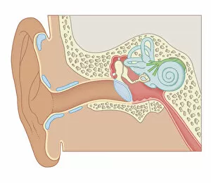
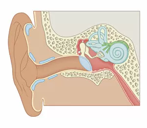
Background imageImages Dated 21st October 2011: Cross section biomedical illustration of internal components of the ear

Cross section biomedical illustration of internal components of the ear

Background imageImages Dated 21st October 2011: Cross section biomedical illustration of human ear

Cross section biomedical illustration of human ear

Background imageImages Dated 21st October 2011: Cross section biomedical illustration of the anatomy of the ear

Cross section biomedical illustration of the anatomy of the ear

Background imageImages Dated 21st October 2011: Cross section biomedical illustration of the anatomy of the ear

Cross section biomedical illustration of the anatomy of the ear

Background imageImages Dated 21st October 2011: Cross section biomedical illustration of partial colectomy

Cross section biomedical illustration of partial colectomy

Background imageImages Dated 21st October 2011: Cross section biomedical illustration of bone repairing itself with clot forming to seal broken

Cross section biomedical illustration of bone repairing itself with clot forming to seal broken blood vessels and fibrous tissue forming to replace the clot

Background imageImages Dated 21st October 2011: Cross section biomedical illustration of bone repairing itself with new, soft

Cross section biomedical illustration of bone repairing itself with new, soft, spongy callus developing on framework provided by fibrous tissue, joining the broken ends

Background imageImages Dated 21st October 2011: Cross section biomedical illustration of bone repairing itself with dense

Cross section biomedical illustration of bone repairing itself with dense, compact bone gradually replacing the callus, and blood vessel regrowth

Background imageImages Dated 21st October 2011: Cross section biomedical illustration of site of incision for microdiscectomy surgical procedure

Cross section biomedical illustration of site of incision for microdiscectomy surgical procedure

Background imageImages Dated 21st October 2011: Cross section biomedical illustration of internal position external fixator used to repair broken

Cross section biomedical illustration of internal position external fixator used to repair broken leg

Background imageImages Dated 21st October 2011: Cross section biomedical illustration of endoscope and tubes inserted in abdomen of adult female

Cross section biomedical illustration of endoscope and tubes inserted in abdomen of adult female during Laparoscopic Surgery

Background imageImages Dated 21st October 2011: Cross section biomedical illustration of nasal / sublingual / buccal route with drugs absorbed

Cross section biomedical illustration of nasal / sublingual / buccal route with drugs absorbed through thin mucus
Cross section biomedical illustration of nasal/sublingual/buccal route with drugs absorbed through thin mucus membrane directly into bloodstream

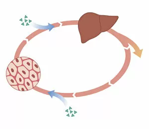
Background imageImages Dated 21st October 2011: Cross section biomedical illustration of how oral and non-oral drugs circulate in the body

Cross section biomedical illustration of how oral and non-oral drugs circulate in the body, showing liver and inset

Background imageImages Dated 21st October 2011: Cross section biomedical illustration of eye cataract surgery sucking softened tissue out using

Cross section biomedical illustration of eye cataract surgery sucking softened tissue out using ultrasound probe inserted into lens via incision in cornea

Background imageImages Dated 21st October 2011: Biomedical illustration of Otoacoustic Emission sound wave chart

Biomedical illustration of Otoacoustic Emission sound wave chart

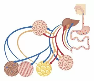
Background imageImages Dated 21st October 2011: Cross section biomedical illustration of how the body uses food for energy, the liver for digestion

Cross section biomedical illustration of how the body uses food for energy, the liver for digestion, fat storage and glycogen storage

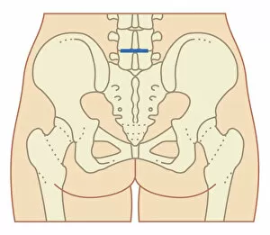
Background imageImages Dated 21st October 2011: Cross section biomedical illustration of rib cage, spine and pelvis, and femur of adult male

Cross section biomedical illustration of rib cage, spine and pelvis, and femur of adult male

Background imageImages Dated 21st October 2011: Cross section biomedical illustration of female anus and rectum

Cross section biomedical illustration of female anus and rectum

Background imageImages Dated 21st October 2011: Cross section biomedical illustration of anatomy during pregnancy

Cross section biomedical illustration of anatomy during pregnancy

Background imageImages Dated 21st October 2011: Cross section biomedical illustration of anatomy at 36 weeks pregnant

Cross section biomedical illustration of anatomy at 36 weeks pregnant

Background imageImages Dated 21st October 2011: Cross section biomedical illustration breast engorgement and sucking position of baby

Cross section biomedical illustration breast engorgement and sucking position of baby

Background imageImages Dated 21st October 2011: Cross section biomedical illustration of anatomy of human eye

Cross section biomedical illustration of anatomy of human eye

Background imageImages Dated 21st October 2011: Cross section biomedical illustration of showing how agonist drugs act on receptors - before

Cross section biomedical illustration of showing how agonist drugs act on receptors - before and after

Background imageImages Dated 21st October 2011: Cross section biomedical illustration of oral route of drugs swallowed

Cross section biomedical illustration of oral route of drugs swallowed and passing into digestive system

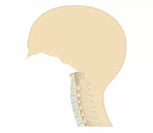
Background imageImages Dated 21st October 2011: Cross section biomedical illustration of vertebral column in neck showing cervical curve

Cross section biomedical illustration of vertebral column in neck showing cervical curve and cervical vertebrae, close-up


All Professionally Made to Order for Quick Shipping


-

Why Choose Us?

We are a leading provider of Art Prints and Photo Gifts since 2004, working in partnership with a range of Sporting Clubs, Charities, Museums and Picture Libraries. A large share of profits from any of their images will go directly towards supporting that charity or club. Our archive of images is carefully curated to bring you a wide range of subjects, including landscapes, wildlife, architecture, and more. We ship from our partner labs in the UK, USA, EU (Netherlands) and Australia.
+

How do I place an order?

Ordering is quick & easy - Just follow 5 Simple Steps:
  1. Find your image: Use our search box or browse our online photo Collections to find the image you want.

  2. Choose your print format: Select your desired product and add it to your cart.

  3. Enter your details: If you're a returning customer, simply enter your email address and password, and we'll fill in your billing and shipping address details. All personal details are held securely and are fully GDPR compliant. As standard, we remove all Personally Identifiable Information after 12 months.

  4. Pay for your purchase: We use state-of-the-art security for online shopping and do not have access to your card details.

  5. Sit back and relax: We'll email you confirmation of your order and when it's dispatched. Registered customers can also track orders in the 'My Account' area.

+

How do I pay for an order?

You can pay for your order with most of the major credit and debit cards, or PayPal. For added security, major financial institutions process payment details separately and securely on our behalf. We do not have access to your online payment card details. Online payments are preferred however we do take phone orders. For UK customers only we accept cheques issued against a UK bank.
+

Is my data safe?

We take data security very seriously. We do not have access to your full card details and all payments are requested over a fully secure connection. Additionally, we fully comply with current European and GDPR legislation, and automatically remove all personal data after at most 12 months (unless you have an account with us and have signed in recently). We also have a strict opt-in policy and would never sell your personal details. Your data is only used to fulfill your order promptly and efficiently.
+

Is wall art ready to hang?

For quick and easy installation all wall art, including framed prints, canvas prints and metal prints are supplied with a ready to hang solution on the back. Generally, saw tooth hangers are applied as they allow wall art to hang flush against the wall. The serrated edge of the hanger prevents the frame from shifting or tilting when hung.
+

Are the photo prints fade resistant?

Yes, we use archival quality photo paper photographic paper for vivid reproduction Prints are an accurate representations of the original artwork, which is preserved for artistic character and authenticity. We guarantee they match previews shown on our web site