Fraud Blocker Skip to main content

Home > All Images > 2010 > April > 14 Apr 2010

Images Dated 14th April 2010 (page 12)

837 items

Background imageImages Dated 14th April 2010: UTAH: STANDING ROCKS. Wood engraving, late 19th century

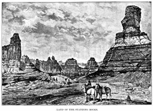
UTAH: STANDING ROCKS. Wood engraving, late 19th century

Background imageImages Dated 14th April 2010: VIEW OF NANTUCKET, 1853. A view of Nantucket. Wood engraving, American, 1853

VIEW OF NANTUCKET, 1853. A view of Nantucket. Wood engraving, American, 1853

Background imageImages Dated 14th April 2010: NANTUCKET, MASSACHUSETTS. A south-eastern view of Nantucket. Wood engraving, American, 1839

NANTUCKET, MASSACHUSETTS. A south-eastern view of Nantucket. Wood engraving, American, 1839

Background imageImages Dated 14th April 2010: CULPEPERs REBELLION. The arrest of George Durant aboard the schooner Carolina on 2 December 1677

CULPEPERs REBELLION. The arrest of George Durant aboard the schooner Carolina on 2 December 1677, the event which sparked Culpepers Rebellion. Line engraving, American, 19th century

Background imageImages Dated 14th April 2010: CALIFORNIA: HACIENDA. Dancing the fandango on a California hacienda in the early 19th century

CALIFORNIA: HACIENDA. Dancing the fandango on a California hacienda in the early 19th century. Line engraving, American, 1891

Background imageImages Dated 14th April 2010: TEXAS: EL PASO, 1860s. El Paso, Texas, in the 1860s. Wood engraving, 1867

TEXAS: EL PASO, 1860s. El Paso, Texas, in the 1860s. Wood engraving, 1867

Background imageImages Dated 14th April 2010: PORT OF HONOLULU, 1849. Wood engraving

PORT OF HONOLULU, 1849. Wood engraving

Background imageImages Dated 14th April 2010: GEORGIA MARKET-PLACE. Marketplace of a Georgia town in cotton season. Wood engraving, 1887

GEORGIA MARKET-PLACE. Marketplace of a Georgia town in cotton season. Wood engraving, 1887

Background imageImages Dated 14th April 2010: ST. JOSEPH, MISSOURI. View of St. Joseph, from the Kansas side of the Missouri River

ST. JOSEPH, MISSOURI. View of St. Joseph, from the Kansas side of the Missouri River: wood engraving, mid-19th century

Background imageImages Dated 14th April 2010: MARBLEHEAD, MASSACHUSETTS Unloading a catch at Marblehead, Massachusetts: wood engraving, 1875

MARBLEHEAD, MASSACHUSETTS Unloading a catch at Marblehead, Massachusetts: wood engraving, 1875

Background imageImages Dated 14th April 2010: TEXAS: THE ALAMO, 1866. Wood engraving from an English newspaper, 1866

TEXAS: THE ALAMO, 1866. Wood engraving from an English newspaper, 1866

Background imageImages Dated 14th April 2010: TOMBSTONE SHERIFF, 1883. The sheriff and his constituents of Tombstone, Arizona. Wood engraving

TOMBSTONE SHERIFF, 1883. The sheriff and his constituents of Tombstone, Arizona. Wood engraving

Background imageImages Dated 14th April 2010: NANTUCKET FISHERMEN, 1875. Outside their homes at Siasconset. Wood engraving, American, 1875

NANTUCKET FISHERMEN, 1875. Outside their homes at Siasconset. Wood engraving, American, 1875

Background imageImages Dated 14th April 2010: TEXAS: RANCHERs CABIN 1875. A ranchers cabin in Texas. Wood engraving from a Missouri Pacific

TEXAS: RANCHERs CABIN 1875. A ranchers cabin in Texas. Wood engraving from a Missouri Pacific Railway guide, c1875
TEXAS:RANCHERs CABIN 1875. A ranchers cabin in Texas. Wood engraving from a Missouri Pacific Railway guide, c1875

Background imageImages Dated 14th April 2010: SC: CHARLESTON, 1739. Detail from a color engraving, 1739

SC: CHARLESTON, 1739. Detail from a color engraving, 1739

Background imageImages Dated 14th April 2010: CALIFORNIA: BIG TREES. The Mariposa Grove of Big Trees (giant sequoias) in the Yosemite Valley

CALIFORNIA: BIG TREES. The Mariposa Grove of Big Trees (giant sequoias) in the Yosemite Valley. Wood engraving, American, 1874

Background imageImages Dated 14th April 2010: Uterus lining, SEM

Uterus lining, SEM
Uterus lining, coloured scanning electron micrograph (SEM). At centre is the opening of a secretory gland. The epithelial cells are covered in uniform short microvilli

Background imageImages Dated 14th April 2010: Smooth muscle, light micrograph

Smooth muscle, light micrograph
Smooth muscle. Light micrograph of a longitudinal section through smooth muscle from the gut. The cells have typically elongated nuclei. Magnification: x240 when printed at 10 centimetres wide

Background imageImages Dated 14th April 2010: Human anatomy, artwork

Human anatomy, artwork
Human anatomy. Computer artwork showing the human nervous, cardiovascular and urinary systems

Background imageImages Dated 14th April 2010: Osteoid osteoma, light micrograph

Osteoid osteoma, light micrograph
Osteoid osteoma. Light micrograph of a section through an osteoid osteoma, a rare benign (non-cancerous) tumour of bone tissue that is very painful

Background imageImages Dated 14th April 2010: Cervical lining, SEM

Cervical lining, SEM
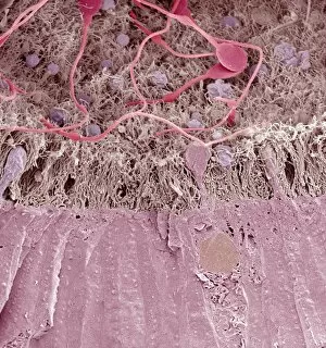
Cervical lining. Coloured scanning electron micrograph (SEM) of microvilli on the lining of the cervix. Magnification: x12000 when printed at 10 centimetres tall

Background imageImages Dated 14th April 2010: Developing pig eye, light micrograph

Developing pig eye, light micrograph. From right the structures seen are: the cornea (brown), the lens (purple), the retina (pink), the choroid (dark brown line) and the sclera (white of the eye)

Background imageImages Dated 14th April 2010: Osteoid osteoma, light micrograph

Osteoid osteoma, light micrograph
Osteoid osteoma. Light micrograph of a section through an osteoid osteoma, a rare benign (non-cancerous) tumour of bone tissue that is very painful

Background imageImages Dated 14th April 2010: Lymphatic system, artwork

Lymphatic system, artwork
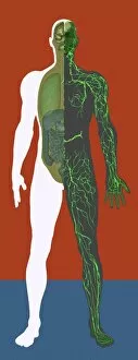
Lymphatic system, computer artwork

Background imageImages Dated 14th April 2010: Human anatomy, artwork

Human anatomy, artwork
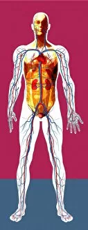
Human anatomy. Computer artwork showing the human urinary and cardiovascular systems

Background imageImages Dated 14th April 2010: Neutrinos, conceptual image

Neutrinos, conceptual image
Neutrinos, conceptual composite image

Background imageImages Dated 14th April 2010: Neutrinos, conceptual image

Neutrinos, conceptual image
Neutrinos, conceptual composite image

Background imageImages Dated 14th April 2010: Smooth muscle, light micrograph

Smooth muscle, light micrograph
Smooth muscle. Light micrograph of a longitudinal section through smooth muscle from the gut. The cells have typically elongated nuclei. Magnification: x240 when printed at 10 centimetres wide

Background imageImages Dated 14th April 2010: Tongue bacteria, SEM

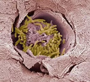
Tongue bacteria, SEM
Tongue bacteria. Coloured scanning electron micrograph (SEM) of rod shaped bacteria (yellow) on the tongue of a Kudu antelope (Tragelaphus sp.). Magnification: x2400 when printed at 10 centimetre wide

Background imageImages Dated 14th April 2010: Hair follicles, light micrograph

Hair follicles, light micrograph
Hair follicles. Light micrograph of a section through skin showing hair follicles. The dermis of the skin is pale blue, the epidermis is green

Background imageImages Dated 14th April 2010: Nervous system, artwork

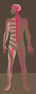
Nervous system, artwork
Nervous system, computer artwork

Background imageImages Dated 14th April 2010: Musculature, artwork

Musculature, artwork
Musculature, computer artwork

Background imageImages Dated 14th April 2010: Cerebellum, light micrograph

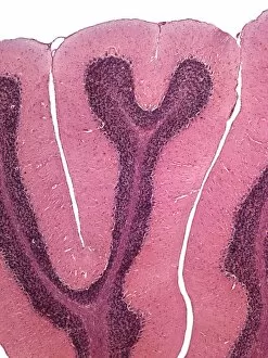
Cerebellum, light micrograph
Cerebellum. Light micrograph of a section through the cerebellum of the brain. The cerebellum comprises three main layers

Background imageImages Dated 14th April 2010: Fat tissue, light micrograph

Fat tissue, light micrograph
Fat tissue. Light micrograph of unilocular adipose (white fat) tissue. Magnification: x160 when printed at 10 centimetres wide

Background imageImages Dated 14th April 2010: Retina, light micrograph

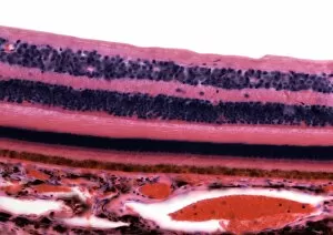
Retina, light micrograph
Retina. Light micrograph of a section through the retina showing its 10 layers. Magnification: x250 when printed at 10 centimetres wide

Background imageImages Dated 14th April 2010: Hair follicles, light micrograph

Hair follicles, light micrograph
Hair follicles. Light micrograph of a section through skin showing hair follicles. The dermis of the skin is pale pink, the epidermis is bright pink

Background imageImages Dated 14th April 2010: Rabbit flea, SEM

Rabbit flea, SEM
Rabbit flea (Spilopsyllus cuniculi), coloured scanning electron micrograph (SEM). The two structures protruding from the centre of the fleas head are sensory palps

Background imageImages Dated 14th April 2010: Immature dog sperm, SEM

Immature dog sperm, SEM
Dog sperm. Coloured scanning electron micrograph (SEM) of immature dog sperm in the epidiymis. These immature sperm are characterised by a small amount of unshed residual cytoplasm about a third of

Background imageImages Dated 14th April 2010: Neutrinos, conceptual image

Neutrinos, conceptual image
Neutrinos, conceptual composite image

Background imageImages Dated 14th April 2010: Human anatomy, artwork

Human anatomy, artwork
Human anatomy. Computer artwork showing the human urinary and cardiovascular systems

Background imageImages Dated 14th April 2010: Skeleton, artwork

Skeleton, artwork
Skeleton, computer artwork

Background imageImages Dated 14th April 2010: Lymphatic system, artwork

Lymphatic system, artwork
Lymphatic system, computer artwork

Background imageImages Dated 14th April 2010: Neutrinos, conceptual image

Neutrinos, conceptual image
Neutrinos, conceptual composite image

Background imageImages Dated 14th April 2010: Fat tissue, light micrograph

Fat tissue, light micrograph
Fat tissue. Light micrograph of unilocular adipose (white fat) tissue. Magnification: x160 when printed at 10 centimetres wide

Background imageImages Dated 14th April 2010: Epididymis, SEM

Epididymis, SEM
Epididymis. Coloured scanning electron micrograph (SEM) of a freeze fracture through the epididymis. Immature sperm (bright pink at top) are seen in the tubule lumen

Background imageImages Dated 14th April 2010: Cervical lining, SEM

Cervical lining, SEM
Cervical lining. Coloured scanning electron micrograph (SEM) of microvilli on the lining of the cervix. Magnification: x12000 when printed at 10 centimetres tall

Background imageImages Dated 14th April 2010: Human anatomy, artwork

Human anatomy, artwork
Human anatomy. Computer artwork showing the human skeleton and cardiovascular, nervous and urinary systems

Background imageImages Dated 14th April 2010: Developing pig eye, light micrograph

Developing pig eye, light micrograph. From right the structures seen are: the cornea (purple), the lens (yellow green), the retina (dark green)


All Professionally Made to Order for Quick Shipping


-

Why Choose Us?

We are a leading provider of Art Prints and Photo Gifts since 2003, working in partnership with a range of Sporting Clubs, Charities, Museums and Picture Libraries. A large share of profits from any of their images will go directly towards supporting that charity or club. Our archive of images is carefully curated to bring you a wide range of subjects, including landscapes, wildlife, architecture, and more. We ship from our partner labs in the UK, USA, EU (Netherlands) and Australia.
+

How do I place an order?

Ordering is quick & easy - Just follow 5 Simple Steps:
  1. Find your image: Use our search box or browse our online photo Collections to find the image you want.

  2. Choose your print format: Select your desired product and add it to your cart.

  3. Enter your details: If you're a returning customer, simply enter your email address and password, and we'll fill in your billing and shipping address details. All personal details are held securely and are fully GDPR compliant. As standard, we remove all Personally Identifiable Information after 12 months.

  4. Pay for your purchase: We use state-of-the-art security for online shopping and do not have access to your card details.

  5. Sit back and relax: We'll email you confirmation of your order and when it's dispatched. Registered customers can also track orders in the 'My Account' area.

+

How do I pay for an order?

You can pay for your order with most of the major credit and debit cards, or PayPal. For added security, major financial institutions process payment details separately and securely on our behalf. We do not have access to your online payment card details. Online payments are preferred however we do take phone orders. For UK customers only we accept cheques issued against a UK bank.
+

Is my data safe?

We take data security very seriously. We do not have access to your full card details and all payments are requested over a fully secure connection. Additionally, we fully comply with current European and GDPR legislation, and automatically remove all personal data after at most 12 months (unless you have an account with us and have signed in recently). We also have a strict opt-in policy and would never sell your personal details. Your data is only used to fulfill your order promptly and efficiently.
+

Is wall art ready to hang?

For quick and easy installation all wall art, including framed prints, canvas prints and metal prints are supplied with a ready to hang solution on the back. Generally, saw tooth hangers are applied as they allow wall art to hang flush against the wall. The serrated edge of the hanger prevents the frame from shifting or tilting when hung.
+

Are the photo prints fade resistant?

Yes, we use archival quality photo paper photographic paper for vivid reproduction Prints are an accurate representations of the original artwork, which is preserved for artistic character and authenticity. We guarantee they match previews shown on our web site