Fraud Blocker Skip to main content

Home > All Images > 2009 > November > 5 Nov 2009

Images Dated 5th November 2009 (page 6)

472 items

Background imageImages Dated 5th November 2009: North America, Mexico

North America, Mexico
Mexico, Guanajuato, San Miguel de Allende, Casa Luna Ranch (Property Released)

Background imageImages Dated 5th November 2009: Mexico, Guanajuato, San Miguel de Allende, Casa Luna Ranch (Property Released)

Mexico, Guanajuato, San Miguel de Allende, Casa Luna Ranch (Property Released)

Background imageImages Dated 5th November 2009: Mexico, Guanajuato, San Miguel de Allende, Casa Luna Ranch (Property Released)

Mexico, Guanajuato, San Miguel de Allende, Casa Luna Ranch (Property Released)

Background imageImages Dated 5th November 2009: John Ignatius Sonny Feehan - Manchester United

John Ignatius Sonny Feehan - Manchester United
John Ignatius Sonny Feehan (Manchester United) 1948 / 49 season. Also Ireland International Photocall. Credit : Colorsport

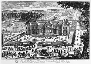
Background imageImages Dated 5th November 2009: CHATEAU DE VAUX LE VICOMTE. The chateau built, 1658-1661, for Nicolas Fouquet

CHATEAU DE VAUX LE VICOMTE. The chateau built, 1658-1661, for Nicolas Fouquet, superintendent of finances of King Louis XIV of France. Contemporary French line engraving

Background imageImages Dated 5th November 2009: Comfrey (Symphytum)

Comfrey (Symphytum)
Foliage, stem and flowers of Comfrey (Symphytum)

Background imageImages Dated 5th November 2009: Ladys Mantle (Alchemilla mollis)

Ladys Mantle (Alchemilla mollis)
Raindrops on foliage of Ladys Mantle (Alchemilla mollis)

Background imageImages Dated 5th November 2009: Black Flies on reeds

Black Flies on reeds
Simuliids on reeds. Known as Black Flies, or Buffalo Gnat, due to the high thorax which gives the fly a hunched appearance. Photographed beside the River Stour in Dorset, UK

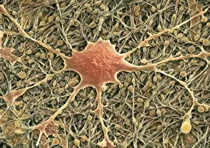
Background imageImages Dated 5th November 2009: Glial cells, SEM

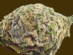
Glial cells, SEM
Glial cells. Coloured scanning electron micrograph (SEM) of a glial cell (centre). Glial cells are nervous system cells that provide structural support and protection for neurons (nerve cells)

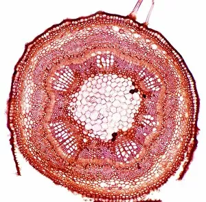
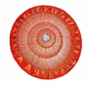
Background imageImages Dated 5th November 2009: Elm stem, light micrograph

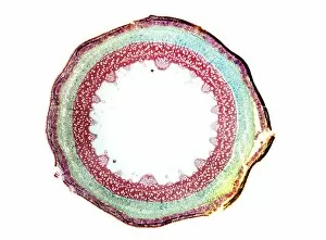
Elm stem, light micrograph
Elm stem. Light micrograph of a transverse section through the one-year-old woody stem of an elm (Ulmus procera) tree. The outer layer (epidermis)

Background imageImages Dated 5th November 2009: Glial cells, SEM

Glial cells, SEM
Glial cells. Coloured scanning electron micrograph (SEM) of a glial cell (centre). Glial cells are nervous system cells that provide structural support and protection for neurons (nerve cells)

Background imageImages Dated 5th November 2009: Nerve cells and glial cells, SEM

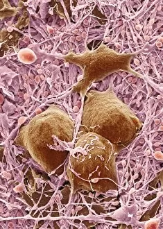
Nerve cells and glial cells, SEM
Nerve cells and glial cells, coloured scanning electron micrograph (SEM). The nerve cells have small cell bodies (green) and fine extensions called axons and dendrites (dark green)

Background imageImages Dated 5th November 2009: Bluebells in woodland

Bluebells in woodland
Bluebells (Hyacinthoides non-scripta) and Hazel (Corylus) in an ancient oak woodland in the spring. Photographed near Wimborne in Dorset, UK

Background imageImages Dated 5th November 2009: Lilac stem, light micrograph

Lilac stem, light micrograph
Lilac stem. Light micrograph of a transverse section through the young woody stem of a lilac (Fraxinus excelsior) tree. The thick epidermis (solid green) is being sloughed (pushed/broken)

Background imageImages Dated 5th November 2009: Nettle Weevil

Nettle Weevil (Phyllobius pomaceus) on a nettle leaf which is common on Nettles between April and September. The golden green colouration comes from thousands of scales, that rub off with wear

Background imageImages Dated 5th November 2009: Field of Cowslips and orchids

Field of Cowslips and orchids
Field of Cowslips (Primula veris) and orchids in flower

Background imageImages Dated 5th November 2009: Nerve cells and glial cells, SEM

Nerve cells and glial cells, SEM
Nerve cells and glial cells, coloured scanning electron micrograph (SEM). The nerve cells have small cell bodies (yellow) and fine extensions called axons and dendrites (pale green)

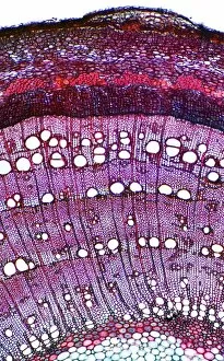
Background imageImages Dated 5th November 2009: Ash stem, light micrograph

Ash stem, light micrograph
Ash stem. Light micrograph of a transverse section through the woody stem of an ash (Fraxinus excelsior) tree showing four growth rings (concentric circles)

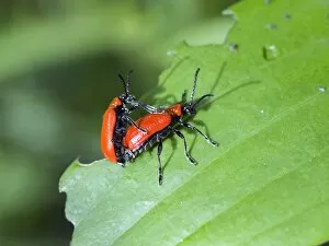
Background imageImages Dated 5th November 2009: Mating Lily Beetles

Mating Lily Beetles (Lilioceris lilii) on a lily (Lilium) leaf

Background imageImages Dated 5th November 2009: Cell infected with HIV, SEM

Cell infected with HIV, SEM
Cell infected with HIV. Coloured scanning electron micrograph (SEM) of HIV particles (pink) budding from the membrane of a host cell

Background imageImages Dated 5th November 2009: Ash stem, light micrograph

Ash stem, light micrograph
Ash stem. Light micrograph of a transverse section through the woody stem of an ash (Fraxinus excelsior) tree showing four growth rings (concentric circles)

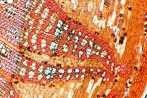
Background imageImages Dated 5th November 2009: Lime tree stem, light micrograph

Lime tree stem, light micrograph
Lime tree stem. Light micrograph of a transverse section through the woody stem of a lime (Tilia cordata) tree. The stem is three-years-old as it has three growth rings (concentric circles)

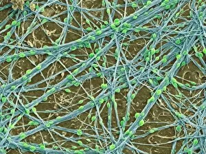
Background imageImages Dated 5th November 2009: Nerve cells, SEM

Nerve cells, SEM
Nerve cells. Coloured scanning electron micrograph (SEM) of nerve cells, showing cell bodies (pink) and extensions called axons and dendrites (purple)

Background imageImages Dated 5th November 2009: Nerve cells and glial cells, SEM

Nerve cells and glial cells, SEM
Nerve cells and glial cells, coloured scanning electron micrograph (SEM). The nerve cells have small cell bodies (blue/pink) and fine extensions called axons and dendrites (blue)

Background imageImages Dated 5th November 2009: Beech (Fagus)

Beech (Fagus)
Spring foliage on Beech trees (Fagus)

Background imageImages Dated 5th November 2009: Nerve cells, SEM

Nerve cells, SEM
Nerve cells. Coloured scanning electron micrograph (SEM) of nerve cells, showing cell bodies (green) and extensions called axons and dendrites (blue)

Background imageImages Dated 5th November 2009: Global temperatures, April 2003

Global temperatures, April 2003. Infrared satellite map of worldwide surface and cloud temperatures in April 2003. The colour-coded map shows temperatures in degrees Celsius

Background imageImages Dated 5th November 2009: Lime tree stem, light micrograph

Lime tree stem, light micrograph
Lime tree stem. Light micrograph of a transverse section through the one-year-old woody stem of a lime (Tilia cordata) tree

Background imageImages Dated 5th November 2009: Clematis montana

Clematis montana
Foliage and flowers of Clematis montana

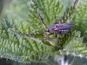
Background imageImages Dated 5th November 2009: Empis Tessellata

Empis Tessellata (Empididae) are true flies (Diptera) with a tiny, spherical head with a down-pointing proboscis and a distinctive short triangular cell close to the wing-tip

Background imageImages Dated 5th November 2009: Beech (Fagus)

Beech (Fagus)
Branches and foliage of Beech (Fagus) in the spring

Background imageImages Dated 5th November 2009: Plant parasite, light micrograph

Plant parasite, light micrograph
Plant parasite. Polarised light micrograph of a transverse section through a plant stem (left) entwined by the common dodder (Cuscuta sp.) parasitic plant (round, right)

Background imageImages Dated 5th November 2009: Aesculus hippocastanum

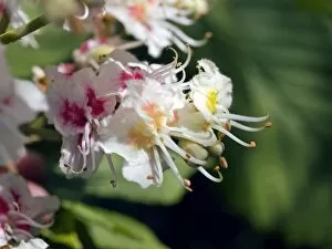
Aesculus hippocastanum
Flowers of Horse Chestnut (Aesculus hippocastanum)

Background imageImages Dated 5th November 2009: Elder tree stem, light micrograph

Elder tree stem, light micrograph
Elder tree stem. Light micrograph of a transverse section through the very young woody stem of an elder (Sambuca nigra) tree

Background imageImages Dated 5th November 2009: Glial cells, SEM

Glial cells, SEM
Glial cells. Coloured scanning electron micrograph (SEM) of a glial cell (centre). Glial cells are nervous system cells that provide structural support and protection for neurons (nerve cells)

Background imageImages Dated 5th November 2009: Cell infected with HIV, SEM

Cell infected with HIV, SEM
Cell infected with HIV. Coloured scanning electron micrograph (SEM) of HIV particles (red/orange) budding from the membrane of a host cell

Background imageImages Dated 5th November 2009: Cell infected with HIV, SEM

Cell infected with HIV, SEM
Cell infected with HIV. Coloured scanning electron micrograph (SEM) of HIV particles (green) budding from the membrane of a host cell

Background imageImages Dated 5th November 2009: Lime tree stem, light micrograph

Lime tree stem, light micrograph
Lime tree stem. Light micrograph of a transverse section through the outer portion of a three-year-old woody stem of a lime (Tilia cordata) tree

Background imageImages Dated 5th November 2009: Nerve cells and glial cells, SEM

Nerve cells and glial cells, SEM
Nerve cells and glial cells, coloured scanning electron micrograph (SEM). The nerve cells have small cell bodies (pink) and fine extensions called axons and dendrites (mauve)

Background imageImages Dated 5th November 2009: Elder tree stem, light micrograph

Elder tree stem, light micrograph
Elder tree stem. Light micrograph of a transverse section through the young woody stem of an elder (Sambuca nigra) tree. The thick epidermis (orange) is being sloughed (pushed/broken)

Background imageImages Dated 5th November 2009: Beech (Fagus)

Beech (Fagus)
Spring foliage on Beech trees (Fagus)

Background imageImages Dated 5th November 2009: Cell infected with HIV, SEM

Cell infected with HIV, SEM
Cell infected with HIV. Coloured scanning electron micrograph (SEM) of HIV particles (red) budding from the membrane of a host cell

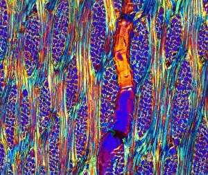
Background imageImages Dated 5th November 2009: Mahogany wood structure, light micrograph

Mahogany wood structure, light micrograph
Mahogany wood structure. Polarised light micrograph of a longitudinal tangential section through a woody stem (xylem) of a mahogany (Swietenia sp.) tree

Background imageImages Dated 5th November 2009: Beech (Fagus)

Beech (Fagus)
Spring foliage on Beech trees (Fagus), showing movement in the wind. The small branches bend and the soft leaves line up with the passing air

Background imageImages Dated 5th November 2009: Nerve cells and glial cells, SEM

Nerve cells and glial cells, SEM
Nerve cells and glial cells, coloured scanning electron micrograph (SEM). The nerve cells have small cell bodies (orange) and fine extensions called axons and dendrites (brown)

Background imageImages Dated 5th November 2009: Nerve cells and glial cells, SEM

Nerve cells and glial cells, SEM
Nerve cells and glial cells, coloured scanning electron micrograph (SEM). The nerve cells have small cell bodies (green) and fine extensions called axons and dendrites (yellow)

Background imageImages Dated 5th November 2009: Global temperatures, May 2009

Global temperatures, May 2009. Infrared satellite map of average daytime air temperatures at 3000 metres elevation worldwide in May 2009

Background imageImages Dated 5th November 2009: Cell infected with HIV, SEM

Cell infected with HIV, SEM
Cell infected with HIV. Coloured scanning electron micrograph (SEM) of HIV particles (green) budding from the membrane of a host cell


All Professionally Made to Order for Quick Shipping


-

Why Choose Us?

We are a leading provider of Art Prints and Photo Gifts since 2004, working in partnership with a range of Sporting Clubs, Charities, Museums and Picture Libraries. A large share of profits from any of their images will go directly towards supporting that charity or club. Our archive of images is carefully curated to bring you a wide range of subjects, including landscapes, wildlife, architecture, and more. We ship from our partner labs in the UK, USA, EU (Netherlands) and Australia.
+

How do I place an order?

Ordering is quick & easy - Just follow 5 Simple Steps:
  1. Find your image: Use our search box or browse our online photo Collections to find the image you want.

  2. Choose your print format: Select your desired product and add it to your cart.

  3. Enter your details: If you're a returning customer, simply enter your email address and password, and we'll fill in your billing and shipping address details. All personal details are held securely and are fully GDPR compliant. As standard, we remove all Personally Identifiable Information after 12 months.

  4. Pay for your purchase: We use state-of-the-art security for online shopping and do not have access to your card details.

  5. Sit back and relax: We'll email you confirmation of your order and when it's dispatched. Registered customers can also track orders in the 'My Account' area.

+

How do I pay for an order?

You can pay for your order with most of the major credit and debit cards, or PayPal. For added security, major financial institutions process payment details separately and securely on our behalf. We do not have access to your online payment card details. Online payments are preferred however we do take phone orders. For UK customers only we accept cheques issued against a UK bank.
+

Is my data safe?

We take data security very seriously. We do not have access to your full card details and all payments are requested over a fully secure connection. Additionally, we fully comply with current European and GDPR legislation, and automatically remove all personal data after at most 12 months (unless you have an account with us and have signed in recently). We also have a strict opt-in policy and would never sell your personal details. Your data is only used to fulfill your order promptly and efficiently.
+

Is wall art ready to hang?

For quick and easy installation all wall art, including framed prints, canvas prints and metal prints are supplied with a ready to hang solution on the back. Generally, saw tooth hangers are applied as they allow wall art to hang flush against the wall. The serrated edge of the hanger prevents the frame from shifting or tilting when hung.
+

Are the photo prints fade resistant?

Yes, we use archival quality photo paper photographic paper for vivid reproduction Prints are an accurate representations of the original artwork, which is preserved for artistic character and authenticity. We guarantee they match previews shown on our web site