Fraud Blocker Skip to main content

Home > All Images > 2009 > July > 17 Jul 2009

Images Dated 17th July 2009 (page 13)

601 items

Background imageImages Dated 17th July 2009: Light Micrograph (LM): A concentration of cells on the epidermis of a plant showing stomata

Light Micrograph (LM): A concentration of cells on the epidermis of a plant showing stomata
LRDS-197 Light Micrograph (LM): A concentration of cells on the epidermis of a plant showing stomata David Spears (Last Refuge)

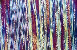
Background imageImages Dated 17th July 2009: Light Micrograph (LM): Longitudinal section shows Scalariform vessels of Fern; Magnification x1200

Light Micrograph (LM): Longitudinal section shows Scalariform vessels of Fern; Magnification x1200 (on 10)
LRDS-196 Light Micrograph (LM): Longitudinal section shows Scalariform vessels of Fern Magnification x1200 (on 10.5 cm width print) David Spears (Last Refuge)

Background imageImages Dated 17th July 2009: Light Micrograph (LM): A longitudinal section f a Ribes sp. stem; Magnification x600 (on 10)

Light Micrograph (LM): A longitudinal section f a Ribes sp. stem; Magnification x600 (on 10. 5 cm width print)
LRDS-195 Light Micrograph (LM): A longitudinal section of a Ribes sp. stem Magnification x600 (on 10.5 cm width print) Ribes sp

Background imageImages Dated 17th July 2009: Light Micrograph (LM): A longitudinal section f a Ribes sp. stem; Magnification x600 (on 10)

Light Micrograph (LM): A longitudinal section f a Ribes sp. stem; Magnification x600 (on 10. 5 cm width print)
LRDS-194 Light Micrograph (LM): A longitudinal section of a Ribes sp. stem Magnification x600 (on 10.5 cm width print) Ribes sp

Background imageImages Dated 17th July 2009: Light Micrograph (LM): A transverse section of a stem of Clubmoss(Lycopodium sp)

Light Micrograph (LM): A transverse section of a stem of Clubmoss(Lycopodium sp. ); Magnification x600 (on 10)
LRDS-185 Light Micrograph (LM): A transverse section of a stem of Clubmoss (Lycopodium sp.) Magnification x600 (on 10.5 cm width print) Lycopodium sp

Background imageImages Dated 17th July 2009: Light Micrograph (LM): transverse section of root of Ranunculus repens (Creeping Buttercup)

Light Micrograph (LM): transverse section of root of Ranunculus repens (Creeping Buttercup)
LRDS-184 Light Micrograph (LM): transverse section of root of Ranunculus repens (Creeping Buttercup) Ranunculus repens David Spears (Last Refuge)

Background imageImages Dated 17th July 2009: Light Micrograph (LM): A transverse section of a stem of Whisk Fern (Psilotum nudum)

Light Micrograph (LM): A transverse section of a stem of Whisk Fern (Psilotum nudum); Magnification x600 (on 10)
LRDS-183 Light Micrograph (LM): A transverse section of a stem of Whisk Fern (Psilotum nudum) Magnification x600 (on 10.5 cm width print) Psilotum nudum David Spears (Last Refuge)

Background imageImages Dated 17th July 2009: Light Micrograph (LM): A transverse section of a stem of Whisk Fern (Psilotum nudum)

Light Micrograph (LM): A transverse section of a stem of Whisk Fern (Psilotum nudum); Magnification x1200 (on 10)
LRDS-182 Light Micrograph (LM): A transverse section of a stem of Whisk Fern Magnification x1200 (on 10.5 cm width print) Psilotum nudum David Spears (Last Refuge)

Background imageImages Dated 17th July 2009: Light Micrograph (LM): transverse section of Prunus Leaf; Magnification x18 (on 10)

Light Micrograph (LM): transverse section of Prunus Leaf; Magnification x18 (on 10. 5 cm width print)
LRDS-181 Light Micrograph (LM): transverse section of Prunus Leaf Magnification x18 (on 10.5 cm width print) Prunus sp David Spears (Last Refuge)

Background imageImages Dated 17th July 2009: Light Micrograph (LM): transverse section shows Sclerenchyma Ground Tissue in Helianthus stem;

Light Micrograph (LM): transverse section shows Sclerenchyma Ground Tissue in Helianthus stem; Magnification x300
LRDS-180 Light Micrograph (LM): transverse section shows Sclerenchyma Ground Tissue in Helianthus stem Magnification x300 (on 10.5 cm width print) Helianthus sp David Spears (Last Refuge)

Background imageImages Dated 17th July 2009: Light Micrograph (LM): transverse section shows Sclerenchyma Ground Tissue in Helianthus stem;

Light Micrograph (LM): transverse section shows Sclerenchyma Ground Tissue in Helianthus stem; Magnification x1200
LRDS-179 Light Micrograph (LM): transverse section shows Sclerenchyma Ground Tissue in Helianthus stem Magnification x1200 (on 10.5 cm width print) Helianthus sp David Spears (Last Refuge)

Background imageImages Dated 17th July 2009: Light Micrograph (LM): transverse section shows Sclerenchyma Ground Tissue in Helianthus stem;

Light Micrograph (LM): transverse section shows Sclerenchyma Ground Tissue in Helianthus stem; Magnification x1200
LRDS-178 Light Micrograph (LM): transverse section shows Sclerenchyma Ground Tissue in Helianthus stem Magnification x1200 (on 10.5 cm width print) Helianthus sp David Spears (Last Refuge)

Background imageImages Dated 17th July 2009: Light Micrograph (LM): A transverse section shows Vascular Bundle in Helianthus stem;

Light Micrograph (LM): A transverse section shows Vascular Bundle in Helianthus stem; Magnification x600 (on 10)
LRDS-177 Light Micrograph (LM): A transverse section shows Vascular Bundle in Helianthus stem Magnification x600 (on 10.5 cm width print) Helianthus sp David Spears (Last Refuge)

Background imageImages Dated 17th July 2009: Light Micrograph (LM): transvers section shows Parenchyma Ground Tissue in Buttercup stem;

Light Micrograph (LM): transvers section shows Parenchyma Ground Tissue in Buttercup stem; Magnification x600 (on 10)
LRDS-176 Light Micrograph (LM): transvers section shows Parenchyma Ground Tissue in Buttercup stem Magnification x600 (on 10.5 cm width print) David Spears (Last Refuge)

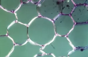
Background imageImages Dated 17th July 2009: Light Micrograph (LM): transverse section shows Parenchyma or packing tissue from Cucurbita Stem;

Light Micrograph (LM): transverse section shows Parenchyma or packing tissue from Cucurbita Stem; Magnification x1200
LRDS-175 Light Micrograph (LM): transverse section shows Parenchyma or packing tissue from Cucurbita Stem Magnification x1200 (on 10.5 cm width print) David Spears (Last Refuge)

Background imageImages Dated 17th July 2009: Light Micrograph (LM): tranverse section shows Parenchyma or packing tissue from Cucurbita Stem;

Light Micrograph (LM): tranverse section shows Parenchyma or packing tissue from Cucurbita Stem; Magnification x600
LRDS-174 Light Micrograph (LM): tranverse section shows Parenchyma or packing tissue from Cucurbita Stem Magnification x600 (on 10.5 cm width print) David Spears (Last Refuge)

Background imageImages Dated 17th July 2009: Light Micrograph (LM): A transverse section of a stem of Clubmoss (Lycopodium sp)

Light Micrograph (LM): A transverse section of a stem of Clubmoss (Lycopodium sp. ); Magnification x600 (on 10)
LRDS-173 Light Micrograph (LM): A transverse section of a stem of Clubmoss (Lycopodium sp.) Magnification x600 (on 10.5 cm width print) David Spears (Last Refuge)

Background imageImages Dated 17th July 2009: Light Micrograph (LM): A transverse section of a stem of Clubmoss (Lycopodium sp)

Light Micrograph (LM): A transverse section of a stem of Clubmoss (Lycopodium sp. ); Magnification x600 (on 10)
LRDS-172 Light Micrograph (LM): A transverse section of a stem of Clubmoss (Lycopodium sp.) Magnification x600 (on 10.5 cm width print) Lycopodium sp

Background imageImages Dated 17th July 2009: Light Micrograph (LM): A transverse section of a stem of Clover (Trifolium sp)

Light Micrograph (LM): A transverse section of a stem of Clover (Trifolium sp. ) with an unidentified parasite attached
LRDS-169 Light Micrograph (LM): A transverse section of a stem of Clover (Trifolium sp.) with an unidentified parasite attached Trifolium sp

Background imageImages Dated 17th July 2009: Light Micrograph (LM): transverse section of Dehiscence Lilium Anther shows Filament

Light Micrograph (LM): transverse section of Dehiscence Lilium Anther shows Filament
LRDS-168 Light Micrograph (LM): transverse section of Dehiscence Lilium Anther shows Filament and Vascular Bundles Magnification x300 (on 10.5 cm width print) David Spears (Last Refuge)


All Professionally Made to Order for Quick Shipping


-

Why Choose Us?

We are a leading provider of Art Prints and Photo Gifts since 2003, working in partnership with a range of Sporting Clubs, Charities, Museums and Picture Libraries. A large share of profits from any of their images will go directly towards supporting that charity or club. Our archive of images is carefully curated to bring you a wide range of subjects, including landscapes, wildlife, architecture, and more. We ship from our partner labs in the UK, USA, EU (Netherlands) and Australia.
+

How do I place an order?

Ordering is quick & easy - Just follow 5 Simple Steps:
  1. Find your image: Use our search box or browse our online photo Collections to find the image you want.

  2. Choose your print format: Select your desired product and add it to your cart.

  3. Enter your details: If you're a returning customer, simply enter your email address and password, and we'll fill in your billing and shipping address details. All personal details are held securely and are fully GDPR compliant. As standard, we remove all Personally Identifiable Information after 12 months.

  4. Pay for your purchase: We use state-of-the-art security for online shopping and do not have access to your card details.

  5. Sit back and relax: We'll email you confirmation of your order and when it's dispatched. Registered customers can also track orders in the 'My Account' area.

+

How do I pay for an order?

You can pay for your order with most of the major credit and debit cards, or PayPal. For added security, major financial institutions process payment details separately and securely on our behalf. We do not have access to your online payment card details. Online payments are preferred however we do take phone orders. For UK customers only we accept cheques issued against a UK bank.
+

Is my data safe?

We take data security very seriously. We do not have access to your full card details and all payments are requested over a fully secure connection. Additionally, we fully comply with current European and GDPR legislation, and automatically remove all personal data after at most 12 months (unless you have an account with us and have signed in recently). We also have a strict opt-in policy and would never sell your personal details. Your data is only used to fulfill your order promptly and efficiently.
+

Is wall art ready to hang?

For quick and easy installation all wall art, including framed prints, canvas prints and metal prints are supplied with a ready to hang solution on the back. Generally, saw tooth hangers are applied as they allow wall art to hang flush against the wall. The serrated edge of the hanger prevents the frame from shifting or tilting when hung.
+

Are the photo prints fade resistant?

Yes, we use archival quality photo paper photographic paper for vivid reproduction Prints are an accurate representations of the original artwork, which is preserved for artistic character and authenticity. We guarantee they match previews shown on our web site