Fraud Blocker Skip to main content

Home > All Images > 2009 > January > 22 Jan 2009

Images Dated 22nd January 2009 (page 6)

369 items

Background imageImages Dated 22nd January 2009: aRON BURR (1756-1836). American political leader. The last days of Aaron Burr

aRON BURR (1756-1836). American political leader. The last days of Aaron Burr. Wood engraving, American, 1892, after a painting, 1889, by Oliver I. Lay

Background imageImages Dated 22nd January 2009: Golf Club

Golf Club
Sean Davies making traditional wooden golf clubs in his workshop at Chulmleigh in Devon

Background imageImages Dated 22nd January 2009: ANCIENT BRITON AND CORACLE. An ancient Briton with his coracle

ANCIENT BRITON AND CORACLE. An ancient Briton with his coracle, as observed by Roman invaders of the 1st century A. D
ANCIENT BRITON AND CORACLE. An ancient Briton with his coracle, as observed by Roman invaders of the 1st century A.D. Line engraving, 19th century

Background imageImages Dated 22nd January 2009: SCOTT: IVANHOE, 1832. Isaac of York and the Templars. Steel engraving from an 1832 English edition

SCOTT: IVANHOE, 1832. Isaac of York and the Templars. Steel engraving from an 1832 English edition of Sir Walter Scotts novel Ivanhoe, first published in 1819

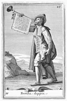
Background imageImages Dated 22nd January 2009: TRUMPET, 1723. Copper engraving, 1723, by Arnold van Westerhout

TRUMPET, 1723. Copper engraving, 1723, by Arnold van Westerhout

Background imageImages Dated 22nd January 2009: ORGAN, 1723. Copper engraving, 1723, by Arnold van Westerhout

ORGAN, 1723. Copper engraving, 1723, by Arnold van Westerhout

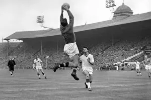
Background imageImages Dated 22nd January 2009: FA Cup Final: Man Utd 2 Aston Villa 1

FA Cup Final: Man Utd 2 Aston Villa 1
Football - 1956 / 1957 FA Cup - Final: Aston Villa 2 Manchester United 1 Villa goalkeeper Nigel Sims and Uniteds Tommy Taylor compete for a ball at Wembley

Background imageImages Dated 22nd January 2009: Peter McParland collides with Ray Wood during the 1957 FA Cup Final

Peter McParland collides with Ray Wood during the 1957 FA Cup Final
Football - 1956 / 1957 FA Cup - Final: Aston Villa 2 Manchester United 1 Ray Wood injury incident: United goakeeper Wood, with ball in hands

Background imageImages Dated 22nd January 2009: FA Cup Final: Man Utd 2 Aston Villa 1

FA Cup Final: Man Utd 2 Aston Villa 1
Football - 1956 / 1957 FA Cup - Final: Aston Villa 2 Manchester United 1 Uniteds Tommy Taylor competes from the ball surrounded by Villa players at Wembley

Background imageImages Dated 22nd January 2009: FA Cup Final: Man Utd 2 Aston Villa 1

FA Cup Final: Man Utd 2 Aston Villa 1
Football - 1956 / 1957 FA Cup - Final: Aston Villa 2 Manchester United 1 Villa goalkeeper Nigel Sims collects a high ball as Uniteds Tommy Taylor, right, closes in, with Uniteds Bobby Charlton, left

Background imageImages Dated 22nd January 2009: FA Cup Final: Man Utd 2 Aston Villa 1

FA Cup Final: Man Utd 2 Aston Villa 1
Football - 1956 / 1957 FA Cup - Final: Aston Villa 2 Manchester United 1 Uniteds Tommy Taylor looks on as Villas Jimmy Dugdale (#5) heads the ball clear at Wembley

Background imageImages Dated 22nd January 2009: Aston Villa - 1957 FA Cup Winners

Aston Villa - 1957 FA Cup Winners
Football - 1956 / 1957 FA Cup - Final: Aston Villa 2 Manchester United 1 Aston Villa captain Johnny Dixon lifts the trophy on his teammates shoulders after victory at Wembley

Background imageImages Dated 22nd January 2009: Nerve fibres, SEM

Nerve fibres, SEM
Nerve fibres. Coloured scanning electron micrograph (SEM) of fractured myelinated nerve fibres. The myelin sheath is blue-green

Background imageImages Dated 22nd January 2009: Vertebra, SEM

Vertebra, SEM
Vertebra. Coloured scanning electron micrograph (SEM) of a single vertebra, part of the vertebral column (spine). The spine is a column of 33 vertebrae (in humans)

Background imageImages Dated 22nd January 2009: Nerve fibres, SEM

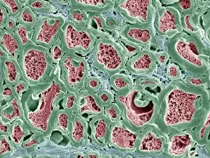
Nerve fibres, SEM
Nerve fibres. Coloured scanning electron micrograph (SEM) of fractured myelinated nerve fibres. The myelin sheath is green, the axoplasm is pink and the endoneurium (connective tissue) is blue

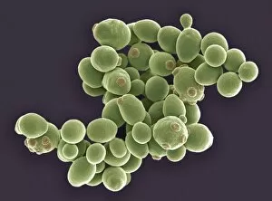
Background imageImages Dated 22nd January 2009: Yeast cells, SEM

Yeast cells, SEM
Yeast cells. Coloured scanning electron micrograph (SEM) of cells of brewers yeast (Saccharomyces cerevisiae). This fungus consists of single vegetative cells

Background imageImages Dated 22nd January 2009: Trachea mucous membrane, SEM

Trachea mucous membrane, SEM
Trachea mucous membrane. Coloured scanning electron micrograph (SEM) of a fractured mucous membrane of the trachea (wind pipe), showing the epithelium and underlying connective tissue

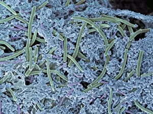
Background imageImages Dated 22nd January 2009: Dental plaque, SEM

Dental plaque, SEM
Dental plaque, coloured scanning electron micrograph (SEM). Plaque consists of bacteria (green and purple) embedded in a glycoprotein matrix (blue)

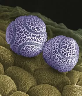
Background imageImages Dated 22nd January 2009: Passion flower pollen, SEM

Passion flower pollen, SEM
Passion flower pollen. Coloured scanning electron micrograph (SEM) of pollen grains from a passion flower (Passiflora caerulea). Pollen grains are the male gametes (sex cells) of a plant

Background imageImages Dated 22nd January 2009: Dental plaque, SEM

Dental plaque, SEM
Dental plaque, coloured scanning electron micrograph (SEM). Plaque consists of bacteria (red and yellow) embedded in a glycoprotein matrix (orange)

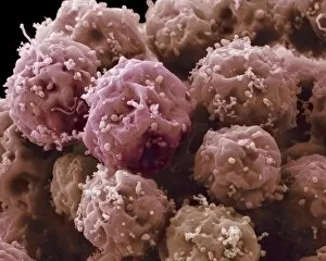
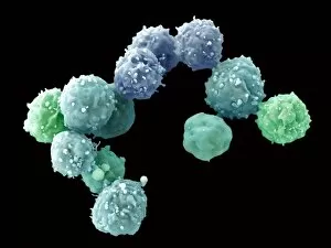
Background imageImages Dated 22nd January 2009: Embryonic stem cells, SEM

Embryonic stem cells, SEM
Embryonic stem cells (ESCs), coloured scanning electron micrograph (SEM). ESCs are pluripotent, that is they are able to differentiate into any cell type

Background imageImages Dated 22nd January 2009: Nerve cell, TEM

Nerve cell, TEM
Nerve cell. Coloured transmission electron micrograph (TEM) of a neurone embedded in grey matter in the central nervous system (CNS)

Background imageImages Dated 22nd January 2009: Embryonic stem cells, SEM

Embryonic stem cells, SEM
Embryonic stem cells (ESCs), coloured scanning electron micrograph (SEM). ESCs are pluripotent, that is they are able to differentiate into any cell type

Background imageImages Dated 22nd January 2009: Yeast cells, SEM

Yeast cells, SEM
Yeast cells. Coloured scanning electron micrograph (SEM) of cells of brewers yeast (Saccharomyces cerevisiae). This fungus consists of single vegetative cells

Background imageImages Dated 22nd January 2009: Nasal lining, TEM

Nasal lining, TEM
Nasal lining. Transmission electron micrograph (TEM) of a section through cilia (green) covering the epithelial lining of the nasopharyx, the part of the throat behind the nasal cavity

Background imageImages Dated 22nd January 2009: Intestinal microvilli, TEM

Intestinal microvilli, TEM
Intestinal microvilli. Coloured transmission electron micrograph (TEM) of a section through microvilli from the small intestine

Background imageImages Dated 22nd January 2009: Smell receptors, TEM

Smell receptors, TEM
Smell receptors. Coloured transmission electron micrograph (TEM) of a section through smell receptors (cilia) projecting from an olfactory neurone (blue)

Background imageImages Dated 22nd January 2009: Embryonic stem cells, SEM

Embryonic stem cells, SEM
Embryonic stem cells (ESCs), coloured scanning electron micrograph (SEM). ESCs are pluripotent, that is they are able to differentiate into any cell type

Background imageImages Dated 22nd January 2009: Pancreas tissue, SEM

Pancreas tissue, SEM
Pancreas tissue. Coloured scanning electron micrograph (SEM) of fractured pancreas tissue. Seen here are zymogen granules (yellow) and cell nuclei (purple)

Background imageImages Dated 22nd January 2009: Nerve fibres, SEM

Nerve fibres, SEM
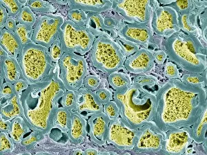
Nerve fibres. Coloured scanning electron micrograph (SEM) of fractured myelinated nerve fibres. The myelin sheath is yellow-green

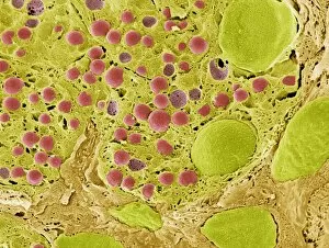
Background imageImages Dated 22nd January 2009: Pancreas tissue, SEM

Pancreas tissue, SEM
Pancreas tissue. Coloured scanning electron micrograph (SEM) of fractured pancreas tissue. Seen here are zymogen granules (red) and cell nuclei (green)

Background imageImages Dated 22nd January 2009: Vertebra, SEM

Vertebra, SEM
Vertebra. Coloured scanning electron micrograph (SEM) of a single vertebra, part of the vertebral column (spine). The spine is a column of 33 vertebrae (in humans)

Background imageImages Dated 22nd January 2009: Nerve fibres, SEM

Nerve fibres, SEM
Nerve fibres. Coloured scanning electron micrograph (SEM) of fractured myelinated nerve fibres. The myelin sheath is blue-green

Background imageImages Dated 22nd January 2009: Trachea mucous membrane, SEM

Trachea mucous membrane, SEM
Trachea mucous membrane. Coloured scanning electron micrograph (SEM) of a fractured mucous membrane of the trachea (wind pipe), showing the epithelium and underlying connective tissue

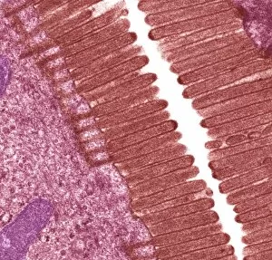
Background imageImages Dated 22nd January 2009: Intestinal microvilli, TEM

Intestinal microvilli, TEM
Intestinal microvilli. Coloured transmission electron micrograph (TEM) of a section through microvilli from the small intestine

Background imageImages Dated 22nd January 2009: Moth proboscis, SEM

Moth proboscis, SEM
Moth proboscis. Coloured scanning electron micrograph (SEM) of the coiled proboscis of a moth (order Lepidoptera). The proboscis is an elongated part of the mouth

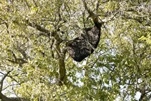
Background imageImages Dated 22nd January 2009: Bee hive in a tree

Bee hive in a tree
Bee hive, covered in bees, in a tree. Photographed in Keoladeo Ghana National Park, Bharatpur, India

Background imageImages Dated 22nd January 2009: Solar water heater

Solar water heater on a rooftop. Solar panels connected to a water tank (white). This produces hot water for domestic use. Photographed in Rajasthan, India

Background imageImages Dated 22nd January 2009: Bacuit archipelago at El Nido, Palawan Island, Philippines, Southeast Asia, Asia

Bacuit archipelago at El Nido, Palawan Island, Philippines, Southeast Asia, Asia

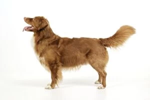
Background imageImages Dated 22nd January 2009: Dog. Nova Scotia Duck Tolling Retriever with tongue out

Dog. Nova Scotia Duck Tolling Retriever with tongue out
JD-21850 Dog. Nova Scotia Duck Tolling Retriever with tongue out John Daniels Please note that prints are for personal display purposes only and may not be reproduced in any way.

Background imageImages Dated 22nd January 2009: Dog. Nova Scotia Duck Tolling Retriever with cups

Dog. Nova Scotia Duck Tolling Retriever with cups
JD-21846 Dog. Nova Scotia Duck Tolling Retriever with cups John Daniels Please note that prints are for personal display purposes only and may not be reproduced in any way.

Background imageImages Dated 22nd January 2009: Dog. Nova Scotia Duck Tolling Retriever

Dog. Nova Scotia Duck Tolling Retriever
JD-21847 Dog. Nova Scotia Duck Tolling Retriever John Daniels Please note that prints are for personal display purposes only and may not be reproduced in any way.

Background imageImages Dated 22nd January 2009: Dog. Nova Scotia Duck Tolling Retriever

Dog. Nova Scotia Duck Tolling Retriever
JD-21848 Dog. Nova Scotia Duck Tolling Retriever John Daniels Please note that prints are for personal display purposes only and may not be reproduced in any way.

Background imageImages Dated 22nd January 2009: Dog. Nova Scotia Duck Tolling Retriever with cups

Dog. Nova Scotia Duck Tolling Retriever with cups
JD-21845 Dog. Nova Scotia Duck Tolling Retriever with cups John Daniels Please note that prints are for personal display purposes only and may not be reproduced in any way.

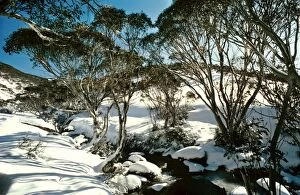
Background imageImages Dated 22nd January 2009: Snow Gums - Kosciuszko National Park, New South Wales, Australia JPF03235

Snow Gums - Kosciuszko National Park, New South Wales, Australia JPF03235
JPF-14125 Snow Gums Kosciuszko National Park, New South Wales, Australia Eucalyptus pauciflora Jean-Paul Ferrero Please note that prints are for personal display purposes only

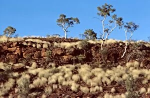
Background imageImages Dated 22nd January 2009: Porcupine Grass and Ghost Gums - Finke Gorge National Park - Northern Territory, Australia JPF03254

Porcupine Grass and Ghost Gums - Finke Gorge National Park - Northern Territory, Australia JPF03254
JPF-14122 Porcupine Grass and Ghost Gums Finke Gorge National Park - Northern Territory, Australia Jean-Paul Ferrero Please note that prints are for personal display purposes only

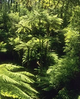
Background imageImages Dated 22nd January 2009: Temperate Rainforest with Rough Tree Ferns - Tarra-Bulga National Park, East Gippsland, Victoria

Temperate Rainforest with Rough Tree Ferns - Tarra-Bulga National Park, East Gippsland, Victoria, Australia JPF03298
JPF-14120 Temperate Rainforest with Rough Tree Ferns Tarra-Bulga National Park, East Gippsland, Victoria, Australia Cyathea australis Jean-Paul Ferrero Please note that prints are for personal

Background imageImages Dated 22nd January 2009: Porcupine Grass and Ghost Gums - Finke Gorge National Park, Northern Territory, Australia JPF03255

Porcupine Grass and Ghost Gums - Finke Gorge National Park, Northern Territory, Australia JPF03255
JPF-14121 Porcupine Grass and Ghost Gums Finke Gorge National Park, Northern Territory, Australia Jean-Paul Ferrero Please note that prints are for personal display purposes only


All Professionally Made to Order for Quick Shipping


-

Why Choose Us?

We are a leading provider of Art Prints and Photo Gifts since 2004, working in partnership with a range of Sporting Clubs, Charities, Museums and Picture Libraries. A large share of profits from any of their images will go directly towards supporting that charity or club. Our archive of images is carefully curated to bring you a wide range of subjects, including landscapes, wildlife, architecture, and more. We ship from our partner labs in the UK, USA, EU (Netherlands) and Australia.
+

How do I place an order?

Ordering is quick & easy - Just follow 5 Simple Steps:
  1. Find your image: Use our search box or browse our online photo Collections to find the image you want.

  2. Choose your print format: Select your desired product and add it to your cart.

  3. Enter your details: If you're a returning customer, simply enter your email address and password, and we'll fill in your billing and shipping address details. All personal details are held securely and are fully GDPR compliant. As standard, we remove all Personally Identifiable Information after 12 months.

  4. Pay for your purchase: We use state-of-the-art security for online shopping and do not have access to your card details.

  5. Sit back and relax: We'll email you confirmation of your order and when it's dispatched. Registered customers can also track orders in the 'My Account' area.

+

How do I pay for an order?

You can pay for your order with most of the major credit and debit cards, or PayPal. For added security, major financial institutions process payment details separately and securely on our behalf. We do not have access to your online payment card details. Online payments are preferred however we do take phone orders. For UK customers only we accept cheques issued against a UK bank.
+

Is my data safe?

We take data security very seriously. We do not have access to your full card details and all payments are requested over a fully secure connection. Additionally, we fully comply with current European and GDPR legislation, and automatically remove all personal data after at most 12 months (unless you have an account with us and have signed in recently). We also have a strict opt-in policy and would never sell your personal details. Your data is only used to fulfill your order promptly and efficiently.
+

Is wall art ready to hang?

For quick and easy installation all wall art, including framed prints, canvas prints and metal prints are supplied with a ready to hang solution on the back. Generally, saw tooth hangers are applied as they allow wall art to hang flush against the wall. The serrated edge of the hanger prevents the frame from shifting or tilting when hung.
+

Are the photo prints fade resistant?

Yes, we use archival quality photo paper photographic paper for vivid reproduction Prints are an accurate representations of the original artwork, which is preserved for artistic character and authenticity. We guarantee they match previews shown on our web site