Fraud Blocker Skip to main content

Home > All Images > 2008 > November > 25 Nov 2008

Images Dated 25th November 2008 (page 6)

423 items

Background imageImages Dated 25th November 2008: Bird feather, light micrograph

Bird feather, light micrograph
Bird feather. Light micrograph of a small contour feather from the body of a bird. This feather, which is 6 millimetres long, is from a juvenile bird

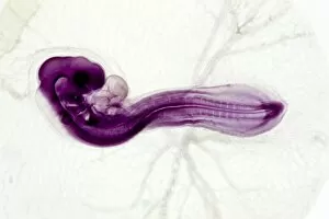
Background imageImages Dated 25th November 2008: Chicken embryo, light micrograph

Chicken embryo, light micrograph
Chicken embryo. Light micrograph of the embryo of a chicken (Gallus domestica), after 60 hours of development, surrounded by the amniotic membrane and yolk blood vessels

Background imageImages Dated 25th November 2008: Human finger, longitudinal section

Human finger, longitudinal section
Human finger. Light micrograph of a longitudinal section through a finger of a human infant. This shows the bones inside the finger (here, the 1st and 2nd phalanges)

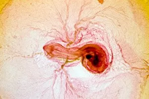
Background imageImages Dated 25th November 2008: Chicken embryo, light micrograph

Chicken embryo, light micrograph
Chicken embryo. Light micrograph of the embryo of a chicken (Gallus domestica) after 3 days of development, surrounded by the amniotic membrane and the yolk blood vessels

Background imageImages Dated 25th November 2008: Tinsley cooling towers demolition

Tinsley cooling towers demolition
Tinsley cooling towers being prepared for demolition. These towers were built in 1938 as part of the Blackburn Meadows power station and stood over 75 metres tall

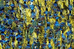
Background imageImages Dated 25th November 2008: Compact bone, light micrograph

Compact bone, light micrograph
Compact bone. Polarised light micrograph of a transverse section through compact bone tissue, showing Haversian canals (circular regions)

Background imageImages Dated 25th November 2008: Dogfish liver area, transverse section

Dogfish liver area, transverse section
Dogfish liver area. Light micrograph of a transverse section through the body of a lesser spotted dogfish (Scyliorhinus canicula), in the region of the liver (lower frame)

Background imageImages Dated 25th November 2008: Woman blowing bubbles

Woman blowing bubbles
MODEL RELEASED. Woman blowing bubbles

Background imageImages Dated 25th November 2008: Tinsley cooling towers, Sheffield

Tinsley cooling towers, Sheffield, UK. These towers were built in 1938 as part of the Blackburn Meadows power station and stood over 75 metres tall

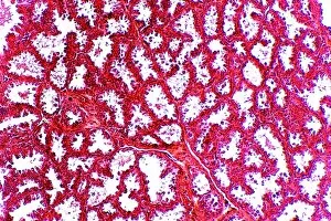
Background imageImages Dated 25th November 2008: Prostate gland, light micrograph

Prostate gland, light micrograph
Prostate gland. Light micrograph of a transverse section through a mammalian prostate gland, an organ found in males that produces fluids to aid the reproductive process

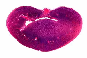
Background imageImages Dated 25th November 2008: Kidney, longitudinal section

Kidney, longitudinal section
Kidney. Light micrograph of a longitudinal section through a kidney, showing the outer region (cortex, round the edges) and the inner region (medulla, centre)

Background imageImages Dated 25th November 2008: Kidney glomerulus, light micrograph

Kidney glomerulus, light micrograph. This section, from the cortex of the kidney, shows the glomerulus (centre) encased in its Bowmans capsule (white arc)

Background imageImages Dated 25th November 2008: Tinsley cooling towers demolition

Tinsley cooling towers demolition
Tinsley cooling towers being demolished. These towers were built in 1938 as part of the Blackburn Meadows power station and stood over 75 metres tall

Background imageImages Dated 25th November 2008: Feather vane, light micrograph

Feather vane, light micrograph
Feather vane. Light micrograph of a part of a vane of a contour feather, from the wing of a bird. Each vane consists of several barbs (black) bearing barbules (thin)

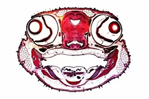
Background imageImages Dated 25th November 2008: Dogfish head, transverse section

Dogfish head, transverse section
Dogfish head. Light micrograph of a transverse section through the head of a lesser spotted dogfish (Scyliorhinus canicula), in the region of the eyes (upper left and upper right)

Background imageImages Dated 25th November 2008: Intervertebral disc, light micrograph

Intervertebral disc, light micrograph
Intervertebral disc. Light micrograph of a transverse section through an intervertebral disc (red) and the surrounding tissue (brown)

Background imageImages Dated 25th November 2008: Heel skin tissue, light micrograph

Heel skin tissue, light micrograph
Heel skin tissue. Polarised light micrograph of a transverse section through skin from the heel of a human foot. The sole of the foot has to withstand the weight of the body

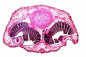
Background imageImages Dated 25th November 2008: Dogfish head, transverse section

Dogfish head, transverse section
Dogfish head. Light micrograph of a transverse section through the head of a lesser spotted dogfish (Scyliorhinus canicula), in the region of the olfactory organs

Background imageImages Dated 25th November 2008: Frog bladder tissue, light micrograph

Frog bladder tissue, light micrograph
Frog bladder tissue. Light micrograph of tissue from the bladder of a frog (Rana temperaria), showing the smooth muscle network (pink)

Background imageImages Dated 25th November 2008: Compact bone, light micrograph

Compact bone, light micrograph
Compact bone. Polarised light micrograph of a transverse section through compact bone tissue, showing Haversian canals (circular regions)

Background imageImages Dated 25th November 2008: Breast tissue, light micrograph

Breast tissue, light micrograph
Breast tissue. Light micrograph of a section through tissue from the human mammary gland during pregnancy. The breast is normally made up of fibro-fatty connective tissue

Background imageImages Dated 25th November 2008: Tinsley cooling towers demolition

Tinsley cooling towers demolition
Tinsley cooling towers being inspected before demolition. These towers were built in 1938 as part of the Blackburn Meadows power station and stood over 75 metres tall

Background imageImages Dated 25th November 2008: Rabbit eye, longitudinal section

Rabbit eye, longitudinal section
Rabbit eye. Light micrograph of a longitudinal section through the eye of a rabbit (Oryctolagus cuniculus). The rabbit has a typical mammalian eye structure

Background imageImages Dated 25th November 2008: Sheeps hoof, light micrograph

Sheeps hoof, light micrograph
Sheeps hoof. Polarised light micrograph of a transverse section through the hoof of a sheep. In sheep and other hooved animals, the hoof is made of the same material as nails and claws

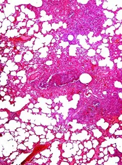
Background imageImages Dated 25th November 2008: Pneumonia infection, light micrograph

Pneumonia infection, light micrograph
Pneumonia infection. Light micrograph of a section through lung tissue from a patient with bronchial pneumonia. Shown here is the blockage and breakdown of the air sacs

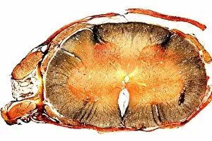
Background imageImages Dated 25th November 2008: Spinal cord, transverse section

Spinal cord, transverse section
Spinal cord. Light micrograph of a transverse section through a human spinal cord. The spinal cord has a central canal around which are the dorsal and ventral horns (orange)

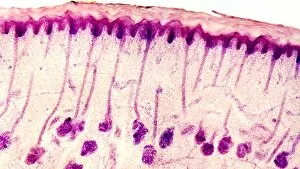
Background imageImages Dated 25th November 2008: Dog tongue tissue, light micrograph

Dog tongue tissue, light micrograph
Dog tongue tissue. Light micrograph of a transverse section through the tongue of a dog, showing the tongues surface (across top)

Background imageImages Dated 25th November 2008: Foot skin tissue, light micrograph

Foot skin tissue, light micrograph
Foot skin tissue. Light micrograph of a transverse section through skin from a human foot. The skin is made up of an outer epidermis (across top)

Background imageImages Dated 25th November 2008: Brain tissue, light micrograph

Brain tissue, light micrograph
Brain tissue. Light micrograph of a section through tissue from the mammalian brain, in this case from the cerebellum. The cerebellum is the part of the brain that controls sensory perception

Background imageImages Dated 25th November 2008: Skin sweat glands, light micrograph

Skin sweat glands, light micrograph
Skin sweat glands. Light micrograph of a transverse section through skin from a human foot. The skin is made up of an outer epidermis (across top) and a lower epidermis (deep purple)

Background imageImages Dated 25th November 2008: Cat tongue tissue, light micrograph

Cat tongue tissue, light micrograph
Cat tongue tissue. Polarised light micrograph of a transverse section through a cats tongue. The outer stratified epithelium (right) has spike-like (white) keratinised papillae

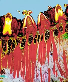
Background imageImages Dated 25th November 2008: Tongue tissue, light micrograph

Tongue tissue, light micrograph
Tongue tissue. Polarised light micrograph of a transverse section through a tongue, showing the surface (across top). Here, there are four rounded fungiform papillae

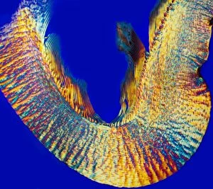
Background imageImages Dated 25th November 2008: Zeolite crystals, light micrograph

Zeolite crystals, light micrograph
Zeolite crystals. Polarised light micrograph of crystals of zeolite from the Giants Causeway, Northern Ireland. Zeolites are hydrated aluminosilicate minerals and have a micro-porous structure

Background imageImages Dated 25th November 2008: Spinal cord, transverse section

Spinal cord, transverse section
Spinal cord. Light micrograph of a transverse section through a mammalian spinal cord. The spinal cord has a central canal around which are the dorsal and ventral horns (deep purple)

Background imageImages Dated 25th November 2008: Skin sweat glands, light micrograph

Skin sweat glands, light micrograph
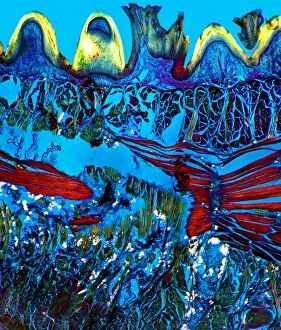
Skin sweat glands. Polarised light micrograph of a transverse section through skin from a human foot. The epidermis (yellow) consists of dead keratinised cells

Background imageImages Dated 25th November 2008: Tongue tissue, light micrograph

Tongue tissue, light micrograph
Tongue tissue. Polarised light micrograph of a transverse section through a tongue, showing the surface (across top). Here, there are three rounded fungiform papillae

Background imageImages Dated 25th November 2008: Liver Building, Liverpool, Merseyside, England

Liver Building, Liverpool, Merseyside, England

Background imageImages Dated 25th November 2008: African Elephant

African Elephant, Loxodonta africana, crossing river in the Masai Mara Game Reserve, Kenya

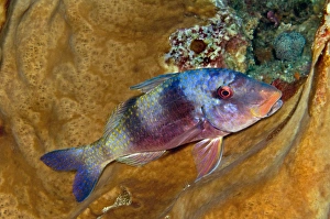
Background imageImages Dated 25th November 2008: Indian Doublebar Goatfish - not usually so colourful this parrot fish has brightened his image

Indian Doublebar Goatfish - not usually so colourful this parrot fish has brightened his image while having a rest in a
VT-9036 Indian Doublebar Goatfish - not usually so colourful this parrot fish has brightened his image while having a rest in a sponge Indonesia Parupeneus trifasciatus Valerie & Ron Taylor Please

Background imageImages Dated 25th November 2008: Ruwenzori Three Horned Chameleon - adult female - Nyungwe - Rwanda - Africa

Ruwenzori Three Horned Chameleon - adult female - Nyungwe - Rwanda - Africa
MM-89 Ruwenzori Three Horned Chameleon - adult female Nyungwe - Rwanda - Africa Chamaeleo jonhstoni Michele Menegon Please note that prints are for personal display purposes only

Background imageImages Dated 25th November 2008: Zitting Cisticola / Streaked Fantail Warbler - perched - Spain

Zitting Cisticola / Streaked Fantail Warbler - perched - Spain
RES-1025 Zitting Cisticola / Streaked Fantail Warbler - perched Spain Cisticola juncidis George Reszeter Please note that prints are for personal display purposes only

Background imageImages Dated 25th November 2008: Zitting Cisticola / Streaked Fantail Warbler - perched - Spain

Zitting Cisticola / Streaked Fantail Warbler - perched - Spain
RES-1024 Zitting Cisticola / Streaked Fantail Warbler - perched Spain Cisticola juncidis George Reszeter Please note that prints are for personal display purposes only

Background imageImages Dated 25th November 2008: Chinese Trumpet Fish - Indo-Pacific: East Africa to Hawaii, north to Japan

Chinese Trumpet Fish - Indo-Pacific: East Africa to Hawaii, north to Japan
PM-10430 Chinese Trumpet Fish Indo-Pacific: East Africa to Hawaii, north to Japan. Eastern and Central Pacific around many islands Aulostomus chinensis Pat Morris Please note that prints are for

Background imageImages Dated 25th November 2008: Upside Down Jellyfish - warm coastal seas, Atlantic and Indian Oceans

Upside Down Jellyfish - warm coastal seas, Atlantic and Indian Oceans
PM-10477 Upside Down Jellyfish warm coastal seas, Atlantic and Indian Oceans. Cassiopea xamachana Pat Morris Please note that prints are for personal display purposes only

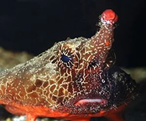
Background imageImages Dated 25th November 2008: Walking Batfish / Toadfish - Caribbean and Gulf of Mexico

Walking Batfish / Toadfish - Caribbean and Gulf of Mexico
PM-10406 Walking Batfish / Toadfish Caribbean and Gulf of Mexico Ogcosephalus nasutus Pat Morris Please note that prints are for personal display purposes only and may not be reproduced in anyway

Background imageImages Dated 25th November 2008: Arapaima - world's largest freshwater fish, Amazon

Arapaima - world's largest freshwater fish, Amazon
PM-10422 Arapaima - world's largest freshwater fish. Arapaima / Pirarucu Fish Amazon. Arapaima gigas Endangered Pat Morris Please note that prints are for personal display purposes only

Background imageImages Dated 25th November 2008: Walking Batfish / Toadfish - Caribbean and Gulf of Mexico

Walking Batfish / Toadfish - Caribbean and Gulf of Mexico
PM-10417 Walking Batfish / Toadfish Caribbean and Gulf of Mexico Ogcosephalus nasutus Pat Morris Please note that prints are for personal display purposes only and may not be reproduced in anyway

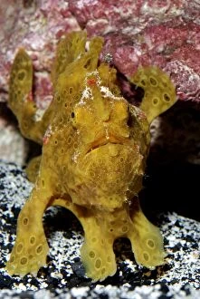
Background imageImages Dated 25th November 2008: Frogfish / Angler Fish - Indo Pacific reefs

Frogfish / Angler Fish - Indo Pacific reefs
PM-10423 Frogfish / Angler Fish - Indo Pacific reefs Antennarius sp. (probably A. pictus) Pat Morris Please note that prints are for personal display purposes only and may not be reproduced in anyway


All Professionally Made to Order for Quick Shipping


-

Why Choose Us?

We are a leading provider of Art Prints and Photo Gifts since 2004, working in partnership with a range of Sporting Clubs, Charities, Museums and Picture Libraries. A large share of profits from any of their images will go directly towards supporting that charity or club. Our archive of images is carefully curated to bring you a wide range of subjects, including landscapes, wildlife, architecture, and more. We ship from our partner labs in the UK, USA, EU (Netherlands) and Australia.
+

How do I place an order?

Ordering is quick & easy - Just follow 5 Simple Steps:
  1. Find your image: Use our search box or browse our online photo Collections to find the image you want.

  2. Choose your print format: Select your desired product and add it to your cart.

  3. Enter your details: If you're a returning customer, simply enter your email address and password, and we'll fill in your billing and shipping address details. All personal details are held securely and are fully GDPR compliant. As standard, we remove all Personally Identifiable Information after 12 months.

  4. Pay for your purchase: We use state-of-the-art security for online shopping and do not have access to your card details.

  5. Sit back and relax: We'll email you confirmation of your order and when it's dispatched. Registered customers can also track orders in the 'My Account' area.

+

How do I pay for an order?

You can pay for your order with most of the major credit and debit cards, or PayPal. For added security, major financial institutions process payment details separately and securely on our behalf. We do not have access to your online payment card details. Online payments are preferred however we do take phone orders. For UK customers only we accept cheques issued against a UK bank.
+

Is my data safe?

We take data security very seriously. We do not have access to your full card details and all payments are requested over a fully secure connection. Additionally, we fully comply with current European and GDPR legislation, and automatically remove all personal data after at most 12 months (unless you have an account with us and have signed in recently). We also have a strict opt-in policy and would never sell your personal details. Your data is only used to fulfill your order promptly and efficiently.
+

Is wall art ready to hang?

For quick and easy installation all wall art, including framed prints, canvas prints and metal prints are supplied with a ready to hang solution on the back. Generally, saw tooth hangers are applied as they allow wall art to hang flush against the wall. The serrated edge of the hanger prevents the frame from shifting or tilting when hung.
+

Are the photo prints fade resistant?

Yes, we use archival quality photo paper photographic paper for vivid reproduction Prints are an accurate representations of the original artwork, which is preserved for artistic character and authenticity. We guarantee they match previews shown on our web site