Fraud Blocker Skip to main content

Home > All Images > 2006 > January > 3 Jan 2006

Images Dated 3rd January 2006 (page 3)

153 items

Background imageImages Dated 3rd January 2006: Copper sulphate crystals

Copper sulphate crystals
Coppersulphate crystals. Polarised light micrograph of copper sulphate (CuS04) crystals

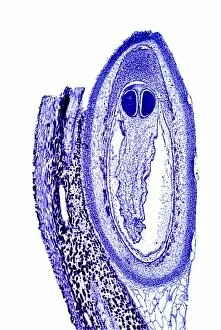
Background imageImages Dated 3rd January 2006: Pine cone scale, light micrograph

Pine cone scale, light micrograph
Pine cone scale. Light micrograph of a vertical section through an ovulate scale from a Scots pine (Pinus sylvestris). This image shows the ovule (oval shape)

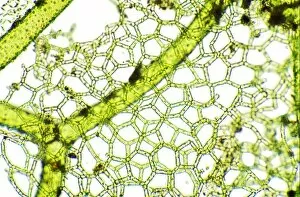
Background imageImages Dated 3rd January 2006: Moss leaf cells, light micrograph

Moss leaf cells, light micrograph
Moss leaf cells. Light micrograph of cells in a leaf of shining Hookeria moss (Hookeria luscens). Two whole cells (horizontal) are seen at centre, surrounded by parts of other cells

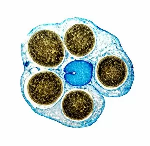
Background imageImages Dated 3rd January 2006: Liverwort spore cases, light micrograph

Liverwort spore cases, light micrograph
Liverwort spore cases. Light micrograph of a cross-section through five spore cases (sporophytes) from the liverwort Conocephalum conicum (Marchantiales)

Background imageImages Dated 3rd January 2006: Liverwort spore cases, light micrograph

Liverwort spore cases, light micrograph
Liverwort spore cases. Light micrograph of a cross-section through five spore cases (sporophytes) from the liverwort Conocephalum conicum (Marchantiales)

Background imageImages Dated 3rd January 2006: Freshwater alga, light micrograph

Freshwater alga, light micrograph
Freshwater alga. Light micrograph of a freshwater alga, the water net Hydrodictyon, showing a full grown net (thick threads) and a young net still growing (thin threads)

Background imageImages Dated 3rd January 2006: Freshwater alga, light micrograph

Freshwater alga, light micrograph
Freshwater alga. Light micrograph of a freshwater alga, the water net Hydrodictyon, showing a full grown net (thick threads) and a young net still growing (thin threads)

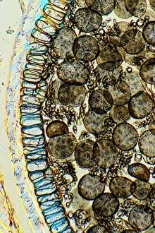
Background imageImages Dated 3rd January 2006: Sporophyte capsule of the liverwort

Sporophyte capsule of the liverwort. Light micrograph of a cross-section through the sporophyte capsule of the liverwort (Conocephalum conicum) common on wet rocks by streams

Background imageImages Dated 3rd January 2006: Abnormal thistle stem, light micrograph

Abnormal thistle stem, light micrograph
Abnormal thistle stem. Light micrograph of a normal (top) and abnormal (bottom) stem of the perennial sow thistle (Sonchus arvensis). The abnormal stem has grown in a flattened, or fasciated, manner

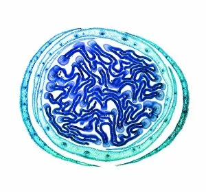
Background imageImages Dated 3rd January 2006: Brazilian liana

Brazilian liana. Light micrograph of the vascular bundles (circular) in the stele of a brazillian liana stem. The red tissue betweent the bundles are medullary rays

Background imageImages Dated 3rd January 2006: Burdock stem, light micrograph

Burdock stem, light micrograph
Burdock stem. Light micrograph of a cross-section through a primary stem of a burdock plant (Arctium lappa). This shows a ring of vascular bundles (red and yellow), which transport water and nutrients

Background imageImages Dated 3rd January 2006: Bladderwort bladder, light micrograph

Bladderwort bladder, light micrograph
Bladderwort bladder. Light micrograph of a bladder (round, lower left) from the greater bladderwort plant (Utricularia vulgaris). A smaller bladder is at centre right. This is a carnivorous plant

Background imageImages Dated 3rd January 2006: Liana stem, light micrograph

Liana stem, light micrograph
Liana stem. Light micrograph of a cross-section through a Brazilian liana stem, showing the large wood vessels (xylem) that channel water

Background imageImages Dated 3rd January 2006: Bracken rhizome, light micrograph

Bracken rhizome, light micrograph
Bracken rhizome. Light micrograph of part of a cross-section through a rhizome from the bracken Pteridium aquilinum. The centre of the rhizome is off top, while the outer surface is off bottom

Background imageImages Dated 3rd January 2006: Stem of a dracaena tree, micrograph

Stem of a dracaena tree, micrograph
Stem of a Dracaena. Light micrograph of a cross- section through the stem of a Dracaena (Darcaena terminalis) of the Agavaceae (Monocotyledons)

Background imageImages Dated 3rd January 2006: Stem of old mans beard

Stem of old mans beard. Light micrograph through the cross section of the stem of old man beard (Clematis vitalba, family Ranunculaceae)

Background imageImages Dated 3rd January 2006: Root of the sago palm, micrograph

Root of the sago palm, micrograph
Sago palm root. Micrograph of a cross-section of the root of the sago palm (Cycas revoluta) showing the diarch xylem and the presence of fungal endotropic mycorrhiza in the cells

Background imageImages Dated 3rd January 2006: Arum plant spadix, light micrograph

Arum plant spadix, light micrograph
Arum plant spadix. Light micrograph of a vertical section through the lower part of the spadix from a lords and ladies (Arum maculatum), or cuckoo pint, plant

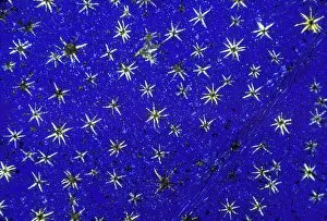
Background imageImages Dated 3rd January 2006: Stellate leaf hairs, light micrograph

Stellate leaf hairs, light micrograph
Stellate leaf hairs. Polarised light micrograph of the epidermis of a leaf of the ornamental shrub Deutzia scabra, showing its stellate (star-shaped) hairs (trichomes, yellow). D

Background imageImages Dated 3rd January 2006: Arum plant spadix, light micrograph

Arum plant spadix, light micrograph
Arum plant spadix. Light micrograph of a vertical section through the spadix from a lords and ladies plant (Arum maculatum)

Background imageImages Dated 3rd January 2006: Leaf bud, light micrograph

Leaf bud, light micrograph
Leaf bud. Light micrograph of a cross-section through a winter leaf bud of the sycamore tree (Acer pseudoplatanus). This trees leaves are arranged in an opposite and decussate manner

Background imageImages Dated 3rd January 2006: Leaf anatomy, light micrograph

Leaf anatomy, light micrograph
Leaf anatomy. Light micrograph of an epidermal peel taken from the surface of a leaf, showing a pattern of epidermal leaf cells and structures, including stomata and their guard cells

Background imageImages Dated 3rd January 2006: Leaf anatomy, light micrograph

Leaf anatomy, light micrograph
Leaf anatomy. Light micrograph of an epidermal peel taken from the surface of a leaf, showing a pattern of leaf veins. An extensive network of small, thin veins is seen between the larger

Background imageImages Dated 3rd January 2006: Leaf bud, light micrograph

Leaf bud, light micrograph
Leaf bud. Light micrograph of a cross-section through a winter leaf bud of the sycamore tree (Acer pseudoplatanus). This trees leaves are arranged in an opposite and decussate manner

Background imageImages Dated 3rd January 2006: Sycamore bud, light micrograph

Sycamore bud, light micrograph
Sycamore bud. Light micrograph of a cross-section through a sycamore bud (Acer pseudo-plantanus). The unexpanded leaves (red) are surrounded by protective outer scale leaves covered in cork

Background imageImages Dated 3rd January 2006: Leaf trichomes

Leaf trichomes. Macrophotograph of the lower surface of a leaf of the plant Elaeagnus pungens, showing the peltate scale hairs or trichomes (lighter colour against dark green of leaf)

Background imageImages Dated 3rd January 2006: Leaf anatomy, light micrograph

Leaf anatomy, light micrograph
Leaf anatomy. Light micrograph of an epidermal peel taken from the surface of a leaf, showing a pattern of epidermal leaf cells and structures, including stomata and their guard cells

Background imageImages Dated 3rd January 2006: Marigold flower parts, light micrograph

Marigold flower parts, light micrograph
Marigold flower parts. Light micrograph of a vertical section through a carpel or pistil (the female part) of a marsh marigold (Caltha palustris)

Background imageImages Dated 3rd January 2006: Leaf anatomy, light micrograph

Leaf anatomy, light micrograph
Leaf anatomy. Light micrograph of an epidermal peel taken from the surface of a leaf, showing a pattern of leaf veins. An extensive network of small, thin veins is seen between the larger

Background imageImages Dated 3rd January 2006: Stellate leaf hairs, light micrograph

Stellate leaf hairs, light micrograph
Stellate leaf hairs. Polarised light micrograph of the epidermis of a leaf of the ornamental shrub Deutzia scabra, showing its stellate (star-shaped) hairs (trichomes, black). D

Background imageImages Dated 3rd January 2006: Copper sulphate and magnesium sulphate

Copper sulphate and magnesium sulphate crystals. Polarised light micrographs of copper sulphate (CuS04) and magnesium sulphate (MgS04) crystals

Background imageImages Dated 3rd January 2006: Silver nitrate crystals

Silver nitrate crystals. Polarised light micrograph of silver nitrate (AgN03) crystals

Background imageImages Dated 3rd January 2006: Fern spore cases, light micrograph

Fern spore cases, light micrograph
Fern spore cases. Polarised light micrograph of a frond of the maidenhair fern (Adiantum capillus- veneris), showing the sporangia (black, centre left) which contain the ferns spores

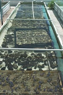
Background imageImages Dated 3rd January 2006: Clam Farm - Trays of Tridacna maxima foreground. On West island Cocos (Keeling) Islands

Clam Farm - Trays of Tridacna maxima foreground. On West island Cocos (Keeling) Islands, Indian Ocean
DH-3702 Clam Farm - Trays of Tridacna maxima foreground On West island Cocos (Keeling) Islands, Indian Ocean. Tridacna maxima Don Hadden Please note that prints are for personal display purposes only

Background imageImages Dated 3rd January 2006: Elder Berries

Elder Berries
ROG-8712 ELDER BERRIES Sambucus nigra Bob Gibbons Please note that prints are for personal display purposes only and may not be reproduced in anyway.

Background imageImages Dated 3rd January 2006: Black Elder

Black Elder
PL-922 Black Elder Sambucus nigra Jens-Peter Laub Please note that prints are for personal display purposes only and may not be reproduced in anyway.

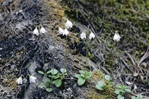
Background imageImages Dated 3rd January 2006: Twin Flower

Twin Flower
JLMO-2549 TWINFLOWER / Twin Flower linnaea borealis John Mason Please note that prints are for personal display purposes only and may not be reproduced in anyway.

Background imageImages Dated 3rd January 2006: Norwegian Mugwort

Norwegian Mugwort
JLM-7282 Norwegian Mugwort / Scottish Wormwood Artemisia norvegica John Mason Please note that prints are for personal display purposes only and may not be reproduced in anyway.

Background imageImages Dated 3rd January 2006: Bogbean

Bogbean
JLM-5074 BOGBEAN / BUCKBEAN - Bog plant, single stem in flower Menyanthes trifoliata Distribution: United Kingdom and Europe

Background imageImages Dated 3rd January 2006: Coconut palm and coconuts - On West Island, Cocos (Keeling) Islands, Indian Ocean

Coconut palm and coconuts - On West Island, Cocos (Keeling) Islands, Indian Ocean
DH-3701 Coconut palm and coconuts - On West Island, Cocos (Keeling) Islands, Indian Ocean. Cocos nucifera Don Hadden Please note that prints are for personal display purposes only

Background imageImages Dated 3rd January 2006: Herb-robert - in autumn

Herb-robert - in autumn
AL-892 HERB Robert - in autumn Geranium robertianum Ake Lindau Please note that prints are for personal display purposes only and may not be reproduced in anyway.

Background imageImages Dated 3rd January 2006: Scarlet Pimpernel Flower

Scarlet Pimpernel Flower
AL-258 Scarlet Pimpernel Flower Anagallis arvensis Ake Lindau Please note that prints are for personal display purposes only and may not be reproduced in anyway.

Background imageImages Dated 3rd January 2006: Mass in Saint-Etienne du Mont church, Paris, France, Europe

Mass in Saint-Etienne du Mont church, Paris, France, Europe

Background imageImages Dated 3rd January 2006: Inca ruins of Pisac, near the village of Pisac, The Sacred Valley, Peru, South America

Inca ruins of Pisac, near the village of Pisac, The Sacred Valley, Peru, South America

Background imageImages Dated 3rd January 2006: View of mainland Antarctica from Danko Island Gentoo penguin colony Antarctica. (RR)

View of mainland Antarctica from Danko Island Gentoo penguin colony Antarctica. (RR)
Juliet Shrimpton / SplashdownDirect

Background imageImages Dated 3rd January 2006: Marine iguana on roadside. Galapagos. (RR)

Marine iguana on roadside. Galapagos. (RR)
Sue Daly / SplashdownDirect

Background imageImages Dated 3rd January 2006: Branched Bur-reed Plant

Branched Bur-reed Plant
ME-676 Branch Bur-Reed Sparganium erectum Johan De Meester Please note that prints are for personal display purposes only and may not be reproduced in any way.

Background imageImages Dated 3rd January 2006: Coconut palms - On a deserted beach on West island, Cocos (Keeling) Islands, Indian Ocean

Coconut palms - On a deserted beach on West island, Cocos (Keeling) Islands, Indian Ocean
DH-3700 Coconut palms - On a deserted beach on West island Cocos (Keeling) Islands, Indian Ocean. Cocos nucifera Don Hadden Please note that prints are for personal display purposes only


All Professionally Made to Order for Quick Shipping


-

Why Choose Us?

We are a leading provider of Art Prints and Photo Gifts since 2004, working in partnership with a range of Sporting Clubs, Charities, Museums and Picture Libraries. A large share of profits from any of their images will go directly towards supporting that charity or club. Our archive of images is carefully curated to bring you a wide range of subjects, including landscapes, wildlife, architecture, and more. We ship from our partner labs in the UK, USA, EU (Netherlands) and Australia.
+

How do I place an order?

Ordering is quick & easy - Just follow 5 Simple Steps:
  1. Find your image: Use our search box or browse our online photo Collections to find the image you want.

  2. Choose your print format: Select your desired product and add it to your cart.

  3. Enter your details: If you're a returning customer, simply enter your email address and password, and we'll fill in your billing and shipping address details. All personal details are held securely and are fully GDPR compliant. As standard, we remove all Personally Identifiable Information after 12 months.

  4. Pay for your purchase: We use state-of-the-art security for online shopping and do not have access to your card details.

  5. Sit back and relax: We'll email you confirmation of your order and when it's dispatched. Registered customers can also track orders in the 'My Account' area.

+

How do I pay for an order?

You can pay for your order with most of the major credit and debit cards, or PayPal. For added security, major financial institutions process payment details separately and securely on our behalf. We do not have access to your online payment card details. Online payments are preferred however we do take phone orders. For UK customers only we accept cheques issued against a UK bank.
+

Is my data safe?

We take data security very seriously. We do not have access to your full card details and all payments are requested over a fully secure connection. Additionally, we fully comply with current European and GDPR legislation, and automatically remove all personal data after at most 12 months (unless you have an account with us and have signed in recently). We also have a strict opt-in policy and would never sell your personal details. Your data is only used to fulfill your order promptly and efficiently.
+

Is wall art ready to hang?

For quick and easy installation all wall art, including framed prints, canvas prints and metal prints are supplied with a ready to hang solution on the back. Generally, saw tooth hangers are applied as they allow wall art to hang flush against the wall. The serrated edge of the hanger prevents the frame from shifting or tilting when hung.
+

Are the photo prints fade resistant?

Yes, we use archival quality photo paper photographic paper for vivid reproduction Prints are an accurate representations of the original artwork, which is preserved for artistic character and authenticity. We guarantee they match previews shown on our web site