Images Dated 21st March 2003
Available as Framed Prints, Photos, Wall Art and Gift Items
Choose from 49 pictures in our Images Dated 21st March 2003 collection for your Wall Art or Photo Gift. Popular choices include Framed Prints, Canvas Prints, Posters and Jigsaw Puzzles. All professionally made for quick delivery.
Abstract
Aerial
Africa
Animals
Architecture
Arts
Asia
Europe
Historic
Humour
Maps and Charts
North America
Oceania
People
Popular Themes
Posters
Religion
Science
Services
South America
Special Days
Sport
Transportation
All Images
> 2003
>> March
>>> 1 Mar 2003
>>> 10 Mar 2003
>>> 11 Mar 2003
>>> 12 Mar 2003
>>> 13 Mar 2003
>>> 14 Mar 2003
>>> 16 Mar 2003
>>> 17 Mar 2003
>>> 18 Mar 2003
>>> 19 Mar 2003
>>> 2 Mar 2003
>>> 20 Mar 2003
>>> 21 Mar 2003
>>> 22 Mar 2003
>>> 23 Mar 2003
>>> 24 Mar 2003
>>> 25 Mar 2003
>>> 26 Mar 2003
>>> 27 Mar 2003
>>> 28 Mar 2003
>>> 3 Mar 2003
>>> 30 Mar 2003
>>> 31 Mar 2003
>>> 4 Mar 2003
>>> 5 Mar 2003
>>> 6 Mar 2003
>>> 7 Mar 2003
>>> 8 Mar 2003
>>> 9 Mar 2003
Collections

Coloured X-ray of an impacted wisdom tooth
Impacted wisdom tooth. Coloured X-ray of teethin the lower left jaw of a patient, showing animpacted wisdom tooth. Normal molar teeth are seenat right (orange) with amalgam fillings in theircrowns (blue). At lower left is a wisdom toothgrowing horizontally, and which has impacted ontothe second molar lying beside it. Adults have fourwisdom teeth (also known as third molars) at theback of each row of teeth. They erupt between theages of 17 and 21. Impacted wisdom teeth maypenetrate the gum incorrectly, or may push otherteeth out of alignment. An impacted wisdom toothsuch as this one may have to be surgicallyremoved
© Alfred Pasieka/Science Photo Library

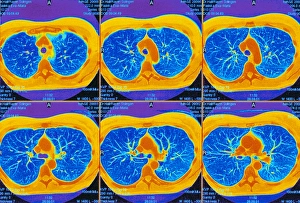
Lungs, CT scans
Lungs. Sequence of six coloured axial CT (computed tomography) scans through the upper chest of a 52- year-old woman. The front of the chest is at top, and the right of the body is at left. The sequence moves down through the body as it runs from left- right along top then bottom. The two lungs (dark blue) and their branching airways (bronchioles, light blue) are separated by the mediastinal area (centre). This contains the trachea (blue circle) that splits into the two lung bronchi (images 4- 6). Major blood vessels (orange, upper centre) lead to the top of the heart (seen in images 4-6). The bone (dark orange) of the spine (lower centre) and ribs (surrounding the lungs) is also seen
© PASIEKA/SCIENCE PHOTO LIBRARY

Spongy bone tissue
Cancellous bone. False-colour scanning electron micrograph of trabeculae in cancellous (spongy) bone tissue. Bone tissue is divided into compact & cancellous. The latter fills the interior of bones & the cavities between the trabeculae are occupied by bone marrow. Trabeculae are thin & composed of irregular lamellae of bone with lacunae containing osteocytes. Some of these lacunae are visible as tiny orange holes at upper centre. Osteblasts are the cells responsible for bone formation. Once they cease activity & become embedded in bone matrix they are called osteocytes. Magnification: x40 at 6x7cm size. Magnification: x40 at 4x5 inch size
© Prof.P.Motta/Dept.Of Anatomy/University la Sapienza , Rome/Science Photo Library

Motorsport Images

Motorsport Images

Motorsport Images

Motorsport Images